
Figure 3.
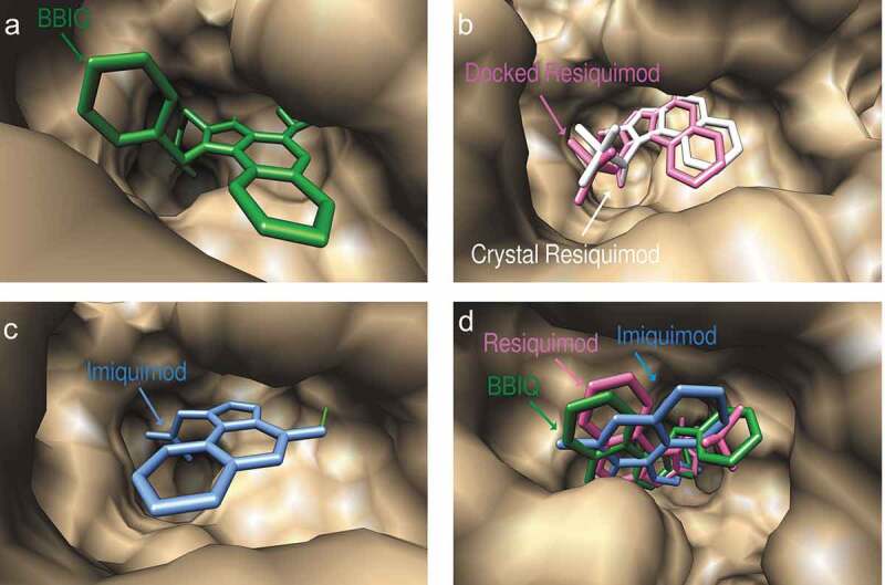
Predicted docked conformations of BBIQ (a), resiquimod (b) and imiquimod (c) to the ligand-binding site of human TLR7, showing predicted hydrogen bonds in green. Figure 3(b) shows a comparison of the predicted and crystal confirmed conformation of resiquimod in the hTLR7 binding pocket with a goodness of fit shown by the Root-mean-square-deviation (RMSD) value of 0.93 Å. Figure 3(d) shows the comparison of the predicted binding poses of BBIQ (in green), resiquimod (in pink) and imiquimod (in blue) in the hTLR7 binding pocket.