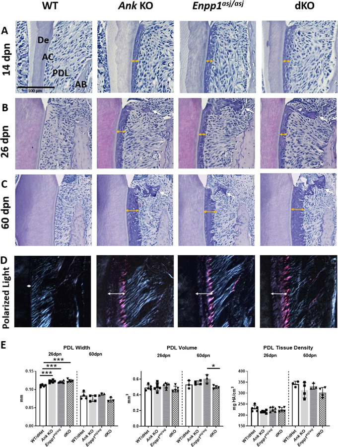
Figure 4. Double Knockout of Ank and Enpp1 Does Not Result in Dental Ankylosis.
At 14dpn (A), 26dpn (B), and 60dpn (C), first molar acellular cementum (AC) width grows to over 10-fold greater thickness vs. WT in Ank and Enpp1asj/asj and dKO mice (double-headed arrows). Cementicles are absent at 14dpn, but present at later stages in the periodontal ligament (PDL) when the root is fully formed (B, C, single-headed white arrows). Microscopic observation of H&E stained sections between crossed polarizers revealed that Ank KO, Enpp1asj/asj, and dKO mice exhibit normal appearing and functionally oriented Sharpey’s fiber insertion into the cementum (D). Quantitative analysis reveals minimal changes in PDL dimensions and no differences in density in Ank KO, Enpp1asj/asj, and dKO mice vs. WT (E). AB: Alveolar bone; De: Dentin.

