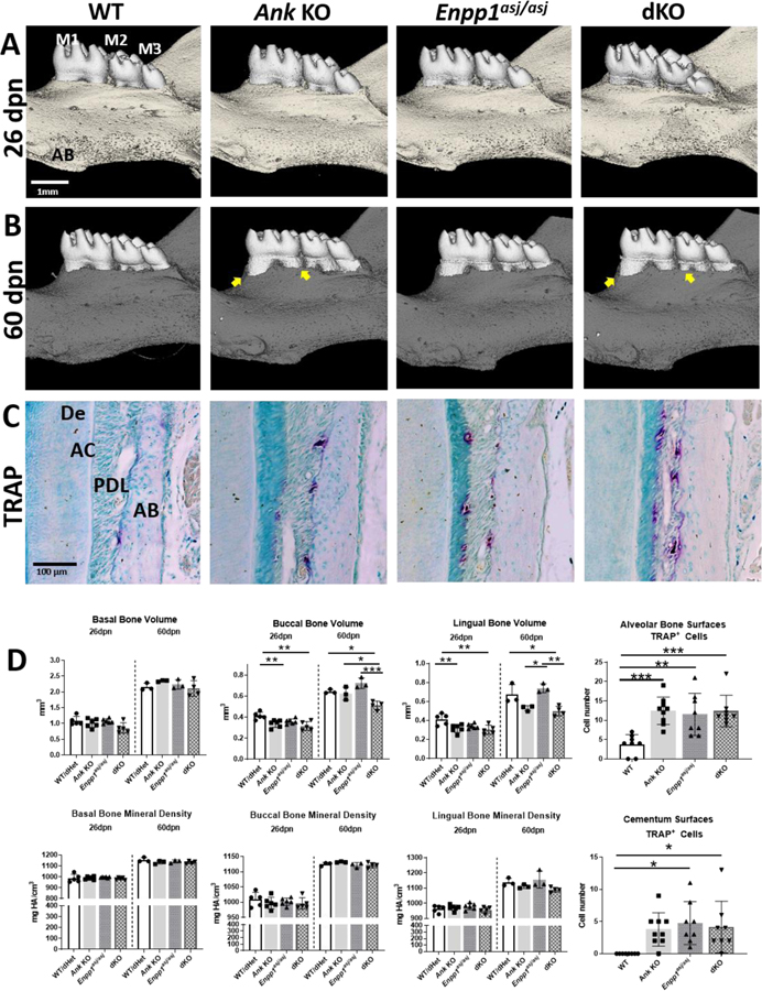
Figure 6. Maintenance of PDL Space by Alveolar Bone Modeling in Mice with Hypercementosis.
3D reconstructions (A, B) of mouse mandibles indicate similar alveolar bone (AB) levels at 26 dpn but AB loss in Ank KO and dKO by 60dpn (B, arrows). TRAP staining (C) reveals osteoclast-like cells (red-purple staining) on alveolar bone and first molar (M1) root surfaces. (D) Quantitative analysis indicates reduced alveolar bone volumes in Ank KO and dKO vs. WT mandibles at 26dpn and/or 60dpn. No differences in alveolar bone density between genotypes are observed. Ank KO, Enpp1asj/asj and dKO mice feature increased numbers of osteoclast-like cells on AB surfaces and odontoclast-like cells on acellular cementum (AC) surfaces. PDL: Periodontal ligament; De: Dentin.

