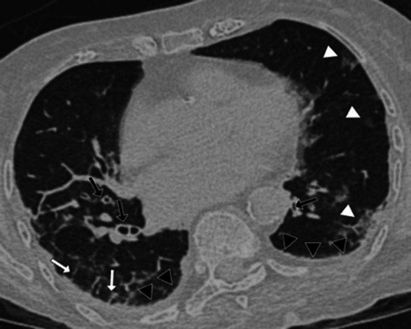
Figure 6. Interstitial and parenchymal findings.
An 88-year-old female patient presented with progressive respiratory distress and equivocal X-ray findings. A chest CT was performed for diagnostic purposes and the subsequent RT-PCR result was positive for COVID-19. An axial unenhanced CT image showed evidence of right bibasal septal thickening (white arrows) with small patchy areas of ground-glass consolidation (white arrowheads). This patient also demonstrated bilateral bronchial wall thickening (black arrows), an unusual finding in our cohort. In addition, there was bilateral basal pleural thickening and shallow pleural effusions (black arrowheads)
COVID-19: coronavirus disease 2019; CT: computed tomography; RT-PCR: reverse transcription-polymerase chain reaction

