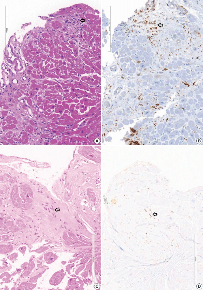
Fig. 1.

Micro-granuloma on the endomyocardial biopsy. (A) Endomyocardial biopsy at 2 years prior to the transplantation of case 1-1 shows confluent fibrosis with edematous stroma. Three foci of infiltration of histiocytes and lymphocytes (arrow) are seen at the margin of fibrosis which is the interface between the fibrosis and myocardium. (B) CD68 staining of the same specimen showing histiocytic infiltration at the micro-granulomas (arrow). (C) Endomyocardial biopsy of case 1-3 shows a micro-granuloma (arrow) of 15 cells in the fibrotic zone. (D) CD68 immunostaining of endomyocardial biopsy of case 1-3 shows positive staining (arrow) on histiocytic marker.
