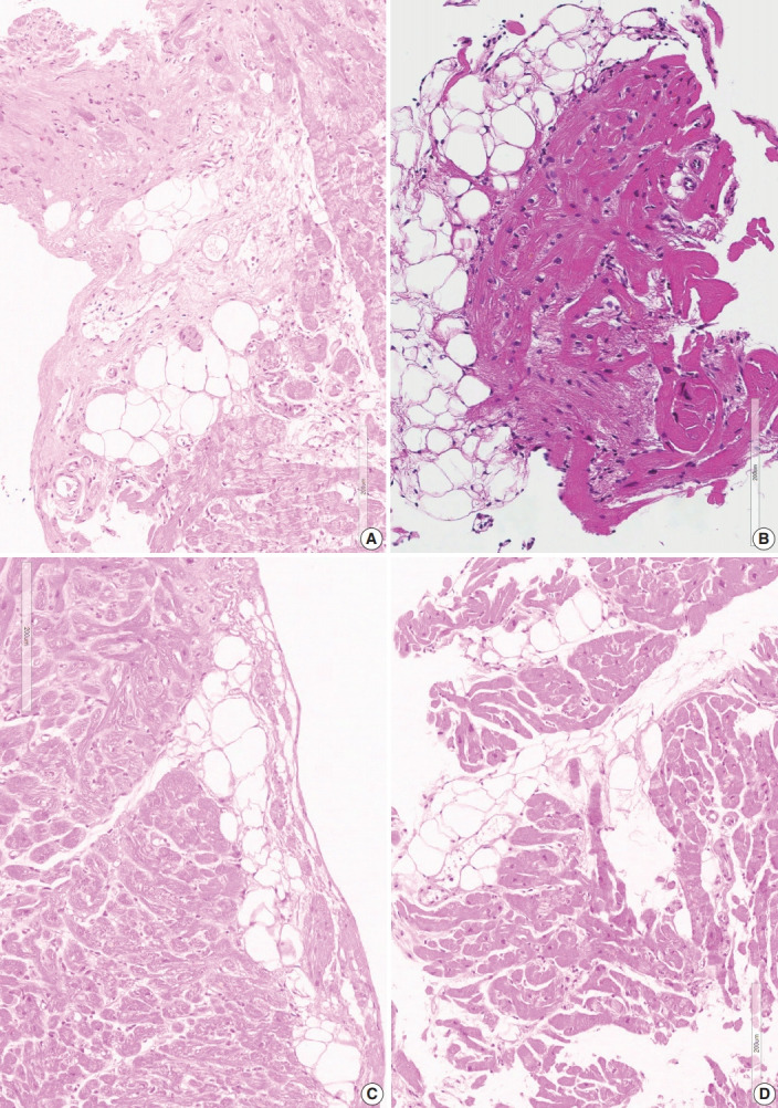
Fig. 4.

Different types of fatty changes in endomyocardial biopsies. (A) Fatty infiltration in the background of confluent fibrosis (case 3-2). (B) Fatty tissue with variable sizes of adipocytes and adjacent myocardium also show post-inflammatory fibrosis (case 1-5). (C) Subendocardial deposition of fatty tissue. Slender fibrotic zone is visible at the margin of fatty area (case 3-5). (D) Fatty infiltration between the myocardial bundles. Adjacent myocardium is normal without fibrosis or inflammation (case 5-4).
