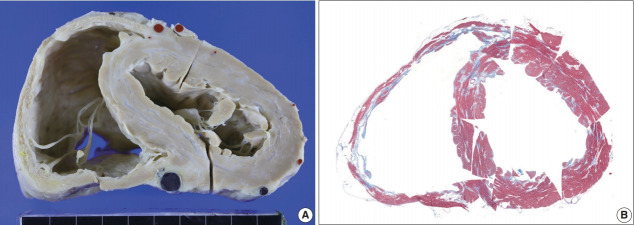
Fig. 6.
A short-axis sectional view of the first transplant heart with sarcoidosis and fibrosis. (A) A short-axis sectional view of the heart shows multifocal confluent fibrosis involving both ventricles. The right ventricular thinning and dilatation are prominent. Coronary arteries and cardiac veins are filled with red and blue silicone rubber cast. (B) Histotopographic mapping of a short-axis plane of the heart by Masson’s trichrome staining reveals prominent fibrosis (in blue color) in the right ventricular free wall and patchy fibrosis in the ventricular septum and left ventricle.

