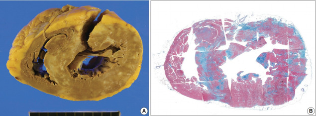
Fig. 7.
The second transplant heart with sarcoidosis and hypertrophied ventricles. (A) Sectional view of the heart shows multifocal confluent fibrosis involving predominantly left ventricle and the ventricular septum. Epicardial fatty tissue is prominent in the right ventricle but the myocardium is not much involved. (B) Histotopographic mapping of a short-axis plane of the heart by Masson’s trichrome staining reveals prominent fibrosis (in blue color) in the interventricular septum and the left ventricular free wall. Distribution of the fibrosis is prominent but not limited to the subepicardial zone.

