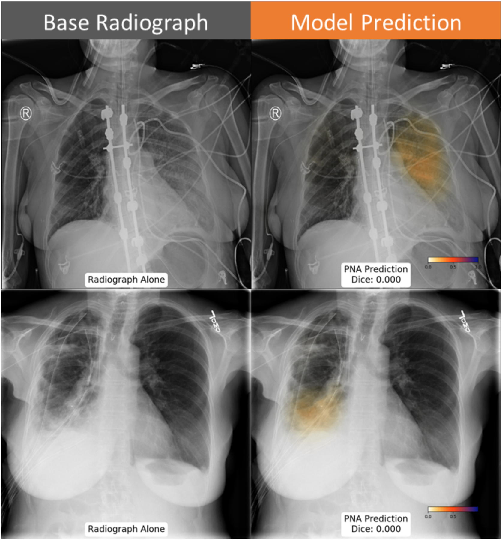
Figure 7: Example cases where neural network predictions disagreed with ground truth annotation.

The neural network predicts pneumonia in the left upper lung (top row), which was not marked by the annotating radiologist. Similarly, the discordant prediction of pneumonia in the right lung in a patient with chest tubes, pleural effusion and adjacent opacity.
