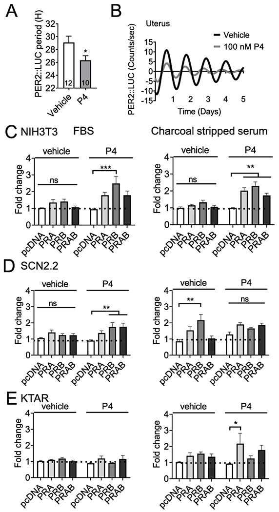
Figure 5. Progesterone regulates Per2-luciferase expression.
A.) Histogram and B.) example trace of PER2::LUC period in the GD 18-19 uterus in response to progesterone (P4 50-100 nM) or veichle control. “PER2::LUC period was analyzed with student’s t-test, N = 10-12, *p < 0.05”. C,-E.) Transient transfections of NIH3T3 (mouse fibroblasts), SCN2.2 (rat SCN cells), and KTAR (mouse arcuate nucleus kisspeptin neurons) with Per2-luciferase, PRA, PRB or empty vector (pcDNA), cultured in in heat inactive heat inactivated FBS (FBS, left side) or charcoal stripped serum (right side). The capacity of progesterone (100 nM) or vehicle to drive Per2-luciferase expression was evaluated. Data is expressed as fold change as compared to control (pcDNA, vehicle). N=3-6 in duplicate. Statistical analysis by two-way ANOVA, followed by a Tukey post hoc. *, p<0.05; **, p<.01; ***, p<.001, ns: non-significant.

