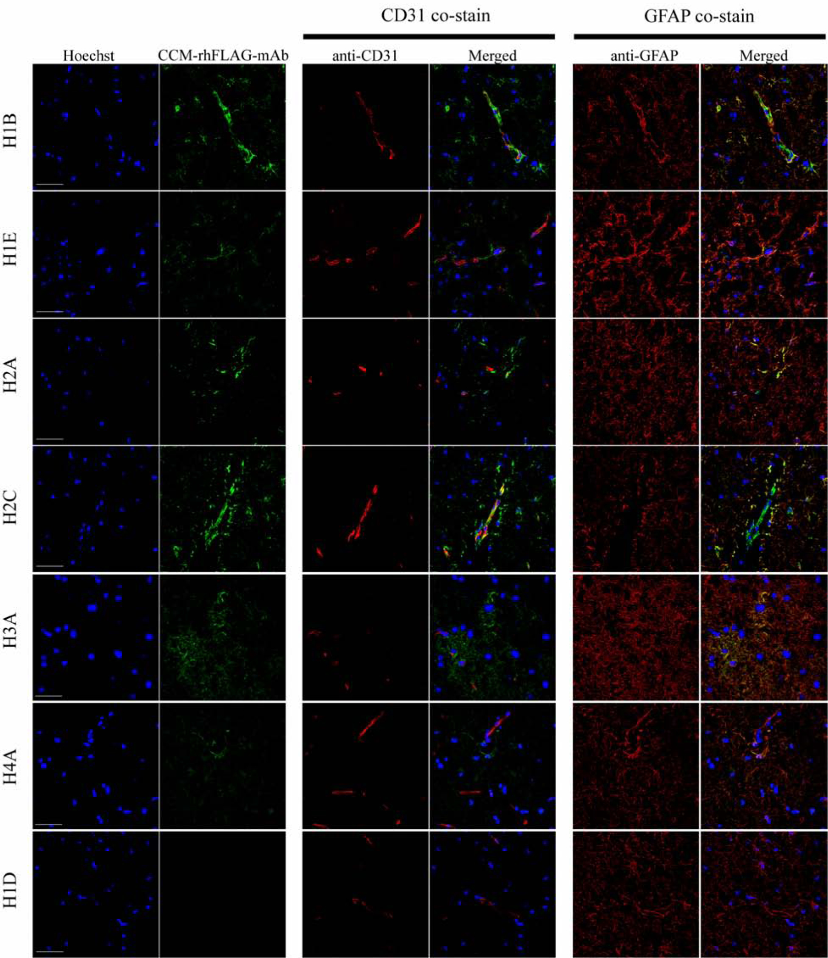
Fig. 2. CCM-rhFLAG-mAbs co-localize with CD31 and GFAP positive cells in normal brain tissue.

Scanning confocal microscopy showing six CCM-rhFLAG-mAb (green, second column from left, top six rows) bound structures within CD31 (red, middle two columns) positive endothelial cell of vessels. The CCM-rhFLAG-mAb (green) bound structures are also found in GFAP (red, right two columns) positive astrocytes adjacent to vessels. Unlike the other six mAbs, H1D CCM-rhFLAG-mAb (bottom row) does not show detectable staining signal. DNA (blue, first column from left) is stained with Hoechst 33342. Scale bars: 50 μm. 2:
