
Figure 1:
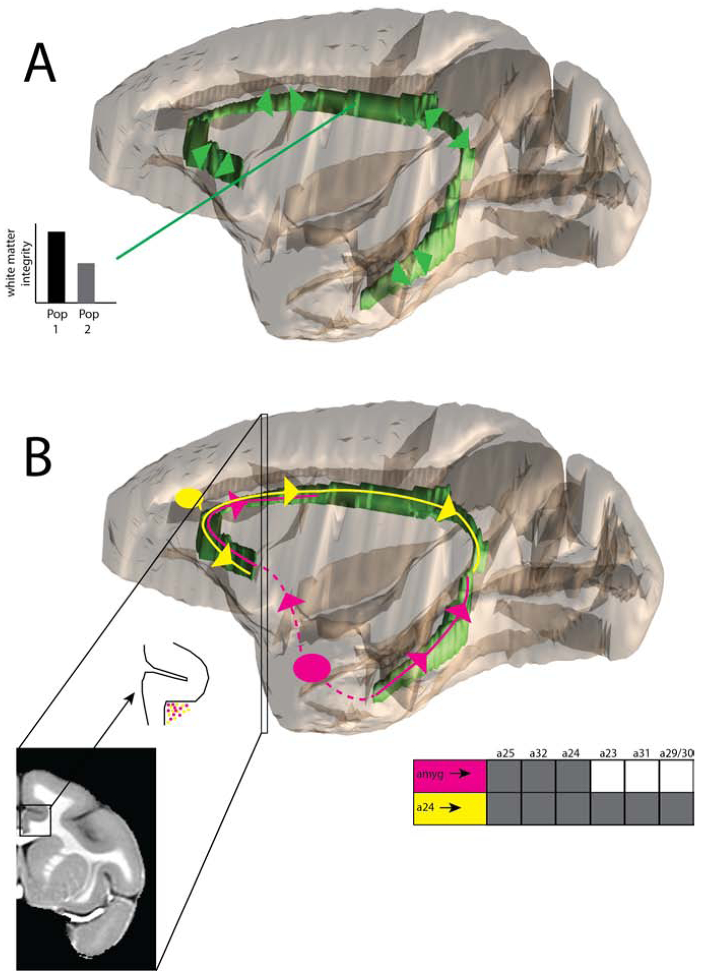
Cartoon illustration of dMRI applications in rhesus macaques, with the cingulum bundle used as an example. A. DMRI can outline the location of a major bundle (cingulum bundle localized in green) as well as the bundle’s major directions (green arrows). This can be used to generate regions-of-interest within the bundle. Metrics, such as fractional anisotropy and apparent diffusion coefficient (discussed in the Introduction), can be compared within these regions-of-interest across populations. B. DMRI is also used to estimate structural connectivity, but this may present challenges. Regions-of-interest are shown in the amygdala (pink) and area 24 (yellow). The true nature of their anterograde projections through the cingulum bundle, as derived from anatomical tract-tracing, are shown as color-matched lines and arrows(88). The amygdala uses the ventral portions of the cingulum bundle (temporal and frontal subgenual), and the rostral portion of the dorsal cingulum bundle. It does not enter the caudal portion of the dorsal cingulum bundle. The resulting anatomical connectivity with anterior cingulate, but not posterior cingulate, is shown as a table, with shaded boxes indicating connectivity(95). By contrast, area 24 uses all of the dorsal cingulum bundle, as well as the frontal subgenual portion of the cingulum bundle. It has anatomical projections to all cingulate regions, as shown by shading in the table(96). It is this anatomical connectivity table that dMRI structural connectivity seeks to re-create, on the basis of streamlines through the white matter. A coronal inset is shown where amygdala and area 24 axons (small colored dots) are both present, and intermixed. A number of challenges are apparent. First, because of overlap with area 24 fibers, it may be challenging to stop amygdala fibers from continuing on to the posterior cingulate cortex. It may also be difficult to correctly identify the points at which fibers from each region join the cingulum bundle. Visualizations created using(97,98).