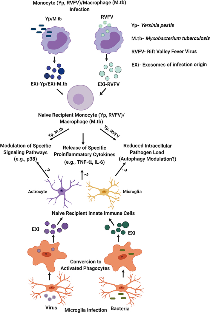
Figure 3: Regulation of innate immune response by exosomes derived from infected monocytes/macrophages (EXi) and potential implications for functions of exosomes produced by infected microglia.
As summarized by the upper half of this figure, the work from our laboratory and published studies from other groups, such as findings for M. tuberculosis infection (Bhatnagar et al. 2007; Singh et al., 2012), have demonstrated that exosomes derived from human monocytes/macrophages infected with bacteria such as Y. pestis or M. tuberculosis, or with the RVFV virus, regulate innate immune responses to infection in specific ways. These include inducing naïve recipient monocytes/macrophages to release proinflammatory cytokines such as TNF-α or IL-6, and inducing significant changes in the activation states of specific signaling proteins in these cells such as the p38 MAP kinase. Our work with both Yp and RVFV has also demonstrated that these exosomes make the naïve recipient cells refractory to subsequent infection, an effect that is likely to involve autophagy since significant changes in autophagy occur as part of host response to both Yp and RVFV (Alem et al. 2015; Moy et al. 2014). The bottom half of this figure summarizes our speculation/hypothesis about microglia, given their role as resident macrophages of the CNS, the findings summarized in the top half of the figure, and the published evidence discussed in the manuscript on effects of exosomes released from infected microglia. We propose that exosomes produced by infected microglia may also have very similar effects on recipient innate immune cells within the CNS (uninfected microglia and astrocytes) as part of mounting an effective response. These may include induction of proinflammatory cytokine release, induction of specific signaling changes, and induction of increased ability for pathogen clearance through modulation of the autophagy pathway.

