Fig. 7.

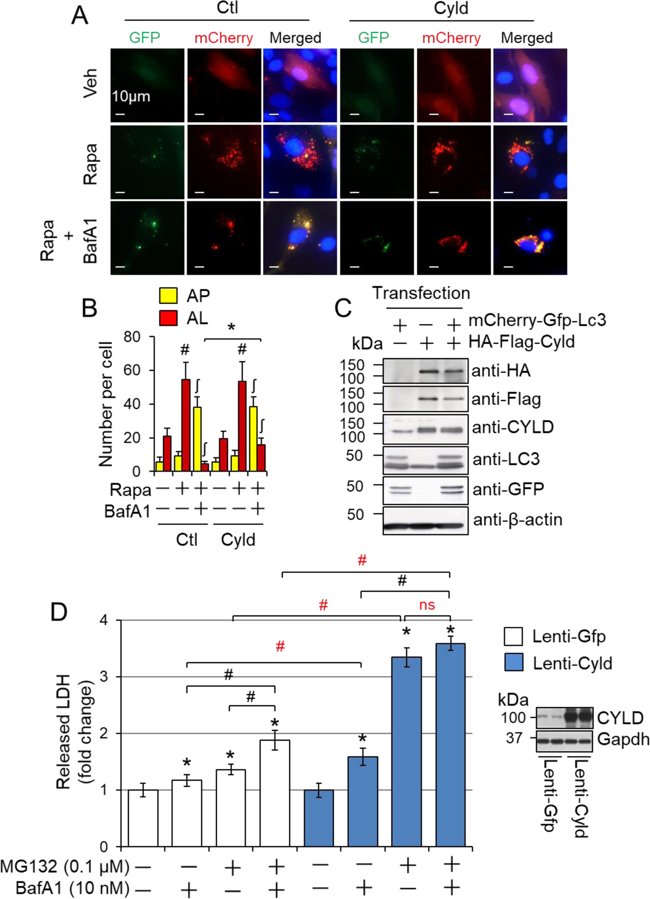
CYLD-mediated suppression of autolysosome clearance and cell death in cardiomyocytes. (A) The representative images of rapamycin (Rapa)-induced autolysosome efflux in H9C2 cells. (B) Quantified numbers of autophagosomes (AP) and autolysosomes (AL). H9C2 cells transfected with mCherry-GFP-LC3 and HA-flag-Cyld plasmids were treated with 1 µM Rapa and 10 nM BafA1 in full growth medium for 24 hours. 50 cells were counted for each group. # or ʃ, p<0.05 vs. vehicle treated controls in the same groups; *, p<0.05 between indicated groups. (C) Verification of transfection efficacy via Western blot analysis for (A) and (B). (D) CYLD overexpression-mediated cell death in H9C2 cells. H9C2 cells stably infected with lentivirus of Gfp (Lenti-Gfp) and lentivirus of Cyld (Lenti-Cyld) were treated with or without MG132 (0.1 µM) and/or BafA1 (10 nM) in serum-free DMEM for 24 hours. The amount of LDH released into supernatants was measured (n=6). The right panel shows the representative immunoblots of CYLD expression in these cells. *, p<0.05 vs. the control (−) in the same group; #, p<0.05 between indicated groups.
