
Fig.3.
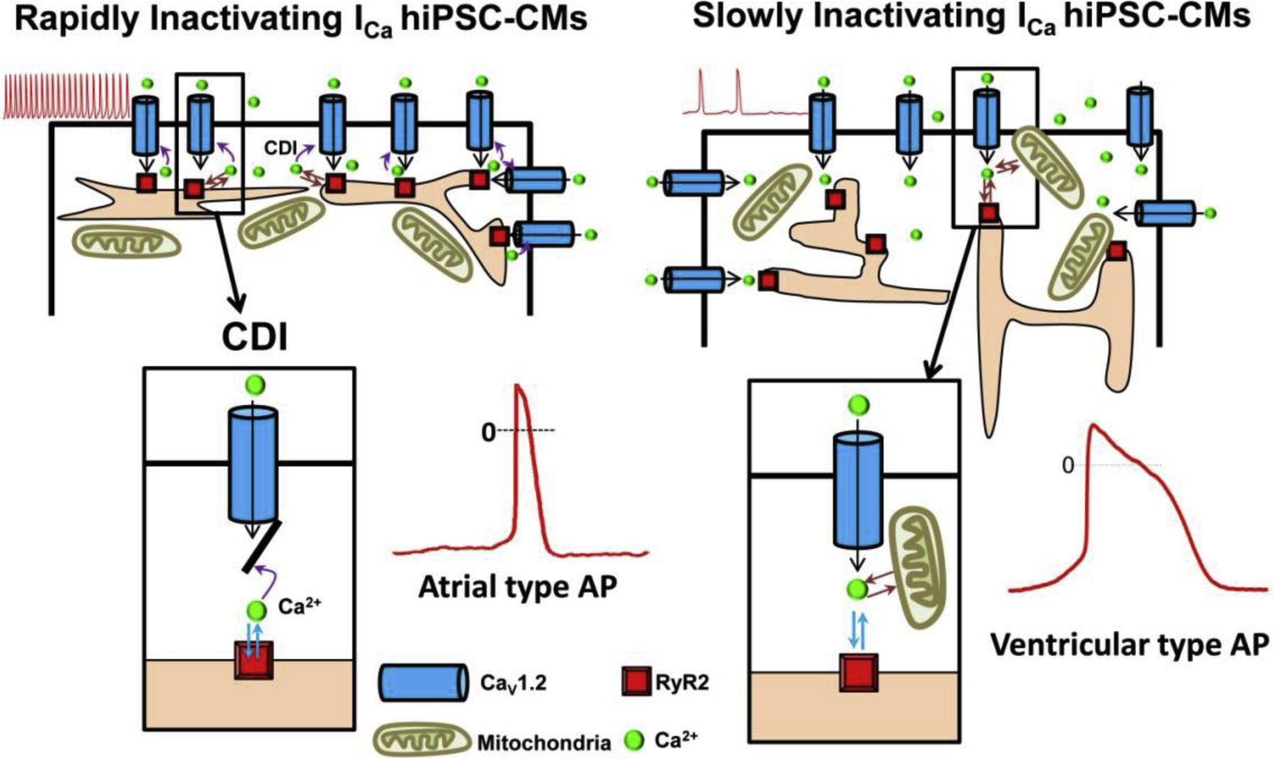
Two cell-type populations of hiPSC-CMs were identified based on the kinetics of ICa inactivation. The 3–4 day post-dissociation smaller hiPSC-CMs, expressed predominantly rapidly inactivating ICa (left panel), shorter action potentials (AP), “atrial-type” and rapid rates of spontaneous activity (red traces) whereas the 6–8 day post-dissociation hiPSC-CMs were larger, expressed mostly slowly inactivating ICa (right panel), longer action potential durations (“Ventricular-type”), and slower rates of spontaneous activity. Enlarged cartoon of calcium dependent inactivation, CDI, responsible for the differential rates of ICa inactivation and durations of APs, showing close proximity of Ca2+ channel with SR release machinery in atrial type APs (left box), and at larger distances in ventricular APs, (right box).