
Fig.5.
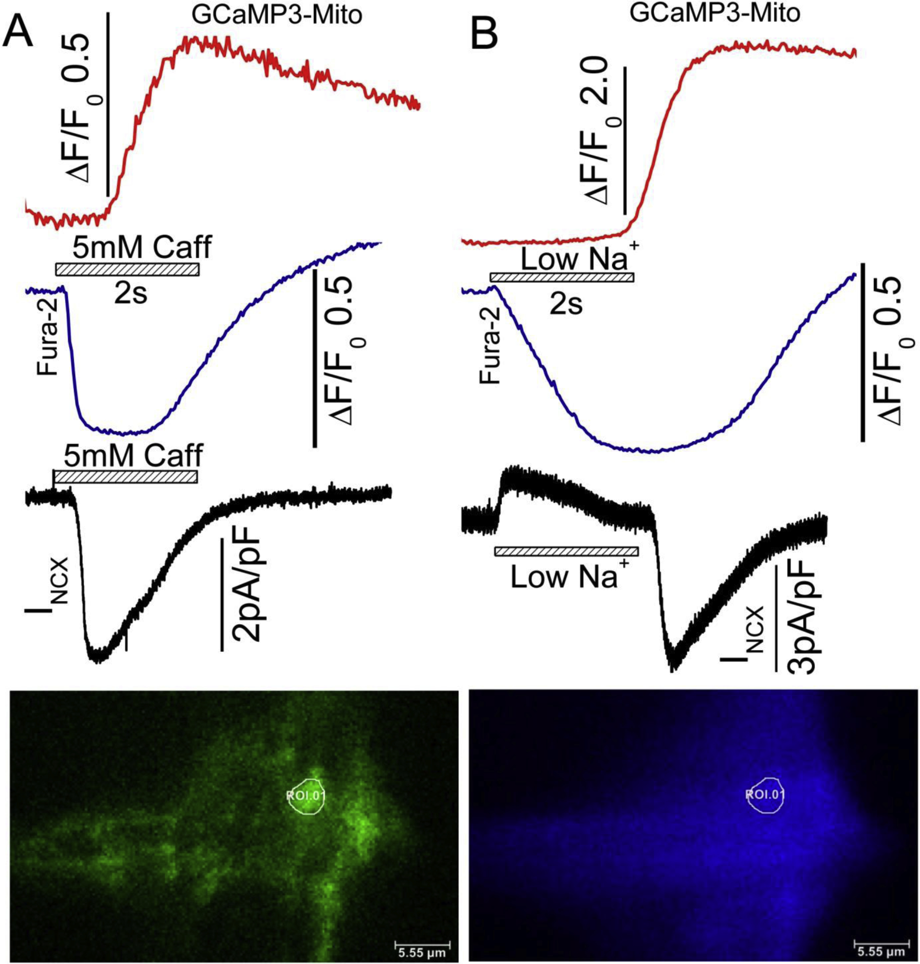
Simultaneously recorded TIRF images of mitochondrial and cytosolic Ca2+ signals in a single patch-clamped hiPSC-CM, in response to Na+ withdrawal (Na+ replaced with TEA) and 5mM caffeine applications. hiPSC-CMs were infected with GCaMP3-mito probe for 72 hours and dialyzed with 0.1mM Fura-2 salt through patch pipet during the experiment. Withdrawal of Na+ activates an outward INCX and its replacement generates a large slowly decaying inward current, that accompanies the rise and fall of cytosolic calcium. The mitochondrial Ca2+ signals recorded from a single mitochondrion (white circle ROI.01 show significant delays when compared to the activation of INCX or rise of cytosolic Ca2+. The blue traces represent cytosolic Fura-2 Ca2+ signal from the same cellular locations. Note that the rise of mitochondrial Ca2+ lags significantly behind the rise in mitochondrial Ca2+. The delays in mitochondrial Ca2+ uptake is shorter on release of SR Ca2+ compared to withdrawal of Na+. The black traces represent the INCX currents activated by Na+ withdraw and caffeine application. GCaMP3-mito probe was excited at 488nm, where increasing fluorescence signal indicates increases in mitochondrial Ca2+. Fura2 was emission signals measured at 405 nm (decreasing fluorescence indicates increase in cytosolic Ca2+). Temperature 35 degrees C.