
Figure 5.
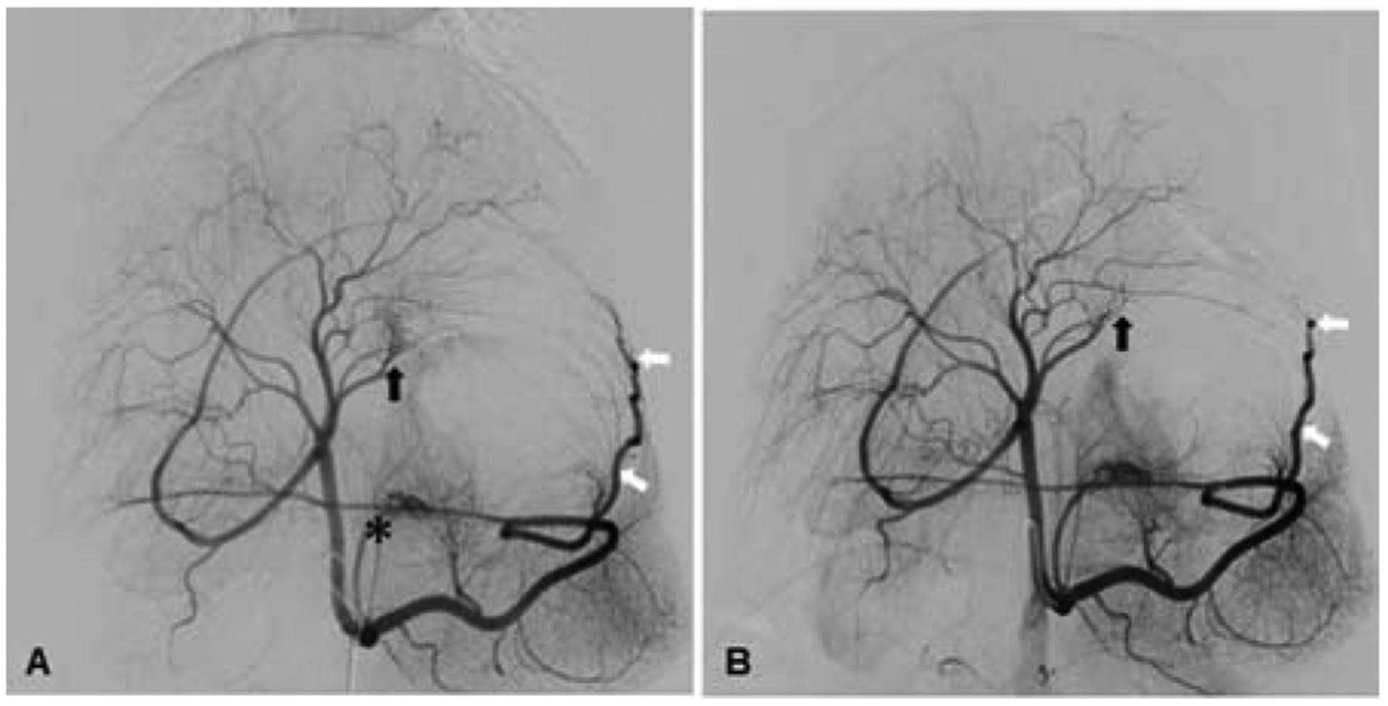
Postmortem identification of XEMs. (A) XEMs were identified in representative tissue blocks from BAE animals by CBCT (arrows). (B) Rhodizonate staining of the tissue sections from BAE animals confirmed the presence of XEMs (arrows).
Official websites use .gov
A
.gov website belongs to an official
government organization in the United States.
Secure .gov websites use HTTPS
A lock (
) or https:// means you've safely
connected to the .gov website. Share sensitive
information only on official, secure websites.
Postmortem identification of XEMs. (A) XEMs were identified in representative tissue blocks from BAE animals by CBCT (arrows). (B) Rhodizonate staining of the tissue sections from BAE animals confirmed the presence of XEMs (arrows).