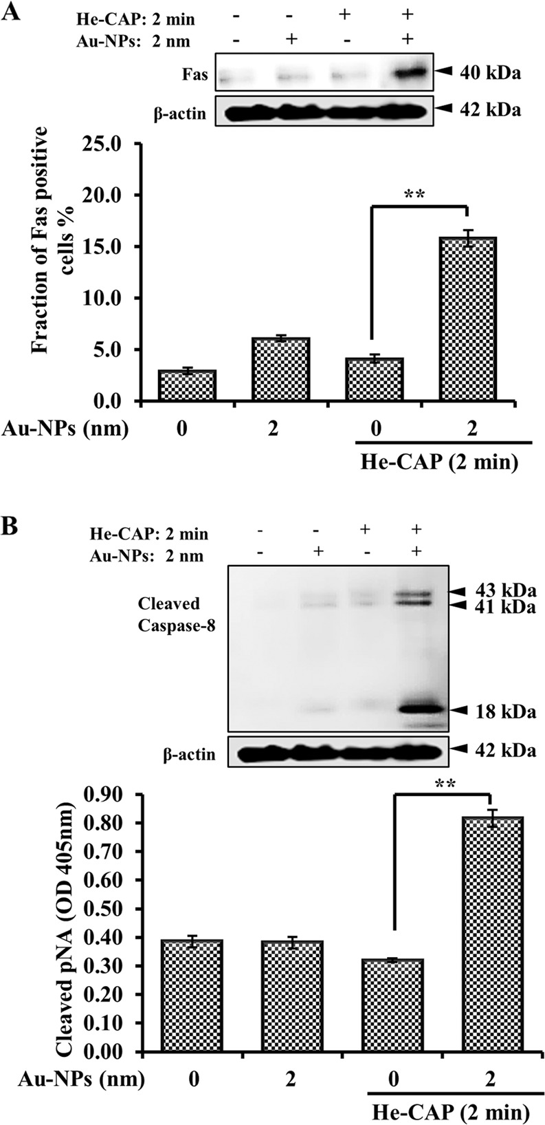
Fig. 5. Effects of Au-NPs on the He-CAP-induced extracellular apoptotic pathway.

a Detection of Fas externalization by western blot analysis and flow cytometry using an anti-Fas FITC-conjugated antibody, 18 h after either treatment. Results are represented as means ± S.E.M. of three independent experiments, **P < 0.001 compared with the He-CAP alone group evaluated by Student’s t test (S.E.M. is indicated by bars). b Caspase-8 activation and expression in the U937 cells were induced by He-CAP alone and in combined treated cells, measured by a western blotting and a FLICE/caspase-8 colorometric protease kit. Results are represented as means ± S.E.M. of three independent experiments, **P < 0.001 compared with the He-CAP alone group evaluated by Student’s t test (S.E.M. is indicated by bars). For western blot β-actin was used to normalize the expression level in each sample. Same β-actin blot was used as the loading control for F.A.S. and cleaved caspase-8 expression. Cropped blots are shown, full-length blots are presented in Supplementary Fig. 3.
