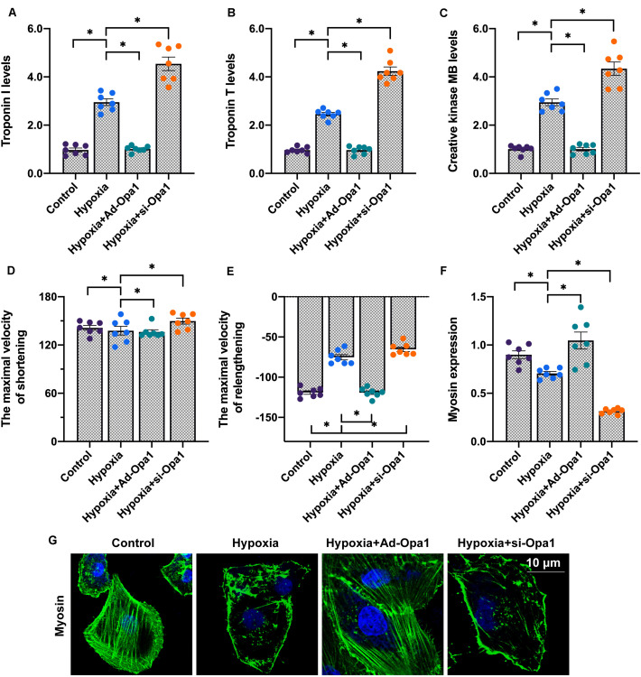
FIGURE 1.
Overexpression of Opa1 attenuates cardiac damage and dysfunction induced by hypoxic stress. (A–C) ELISA analysis of troponin T (TnT), troponin I (TnI), and creatine kinase MB (CK-MB) secretion by Opa1-overexpressing (Ad-Opa1) and Opa1-knockdown (si-Opa1) cardiomyocytes subjected to 24 h hypoxia exposure. (D,E) Analysis of cardiomyocyte contractile properties. Maximal shortening and relengthening velocities were measured using a SoftEdge MyoCam system. (F,G) Myosin immunofluorescence results. ∗p < 0.05.

