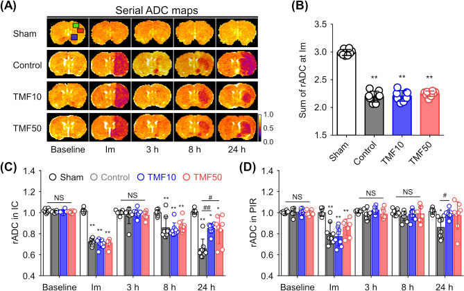
Figure 2.
Effect of TMF treatment on apparent diffusion coefficient (ADC) values (A) Mean ADC values were measured in the ipsilateral ischaemic core (IC, red square), peri-infarct region (PIR, green square), anterior choroidal and hypothalamic region (AHR, blue square), and contralateral regions on ADC maps during the follow-up period. (B) Combined relative ADC (rADC) values immediately after ischaemia were decreased in groups with induced ischaemia. (C,D) The rADC values of the (C) IC and (D) PIR were plotted against time. Data are represented as means ± SDs (n = 8 rats in each group). *P < 0.05 vs. sham group; **P < 0.001 vs. sham group; #P < 0.05 vs. control group; ##P < 0.001 vs. control group.

