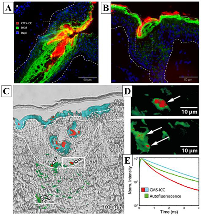
Figure 1.

Example of conventional fluorescence, FLIM and transmitted light images of a drug (DXM) and its core-multishell nanocarrier (CMS-NC) distributions within ex vivo human skin. A / B) conventional fluorescence images highlighting in red the nanocarrier CMS-ICC (indocarbocyanine-labelled CMS-NC), in green the DXM drug (anti-DXM primary-antibody coupled with Fluorescein isothiocyanate (FITC)-labelled secondary-antibody) and in blue the cells nuclei (DAPI staining); C) Transmitted light and conventional FLIM overlay images of CMS-ICC penetration in the skin section. Pseudo-colored fluorescence decay signatures shown in (E) indicate the localization of the CMS-ICC-NC; dashed lines indicate borders between stratum corneum, living epidermis and dermis layers. (D) Cluster-FLIM images of epidermal and dermal ICC-CMS-NC locations; zoom into regions of (C) as indicated by white rectangles. (E) Fluorescence decay signatures (ICC-CMS-NC: red, cyan; Autofluorescence: green) revealed by cluster-FLIM analysis. Reprinted from J. Control. Release, 299, Frombach et al., Core-multishell nanocarriers enhance drug penetration and reach keratinocytes and antigen-presenting cells in intact human skin, 138-148, Copyright (2019), with permission from Elsevier [16].
