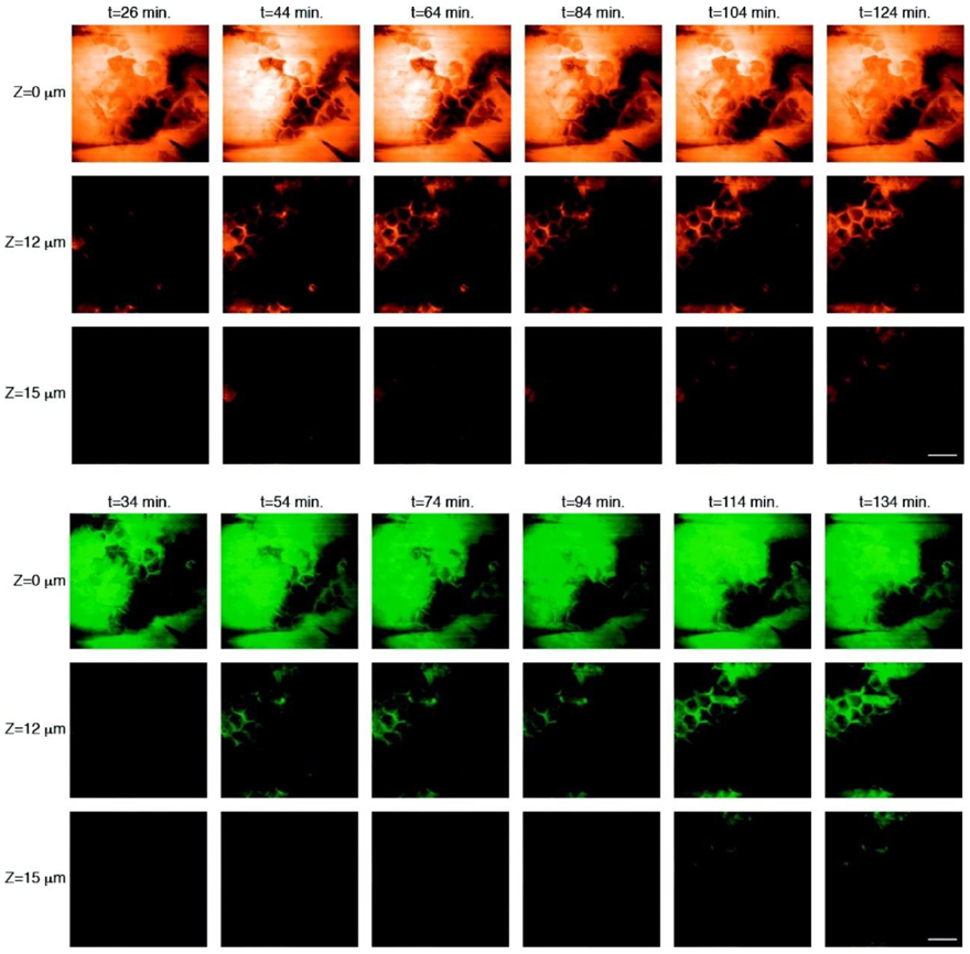
Figure 10.

Imaging the penetration of deuterated PG (upper panel) and ketoprofen (lower panel) across the stratum corneum. Images acquired at the depths indicated down the left-hand side of the figure and times indicated along the top show the penetration of cosolvait and drug into the tissue using SRS contrast at 2120 cm−1 and 1599 cm−1, respectively. Scale bar = 50 μm. Reprinted with permission from Brian et al. Imaging Drug Delivery to Skin with Stimulated Raman Scattering Microscopy, Molecular Pharmaceutics 2011 8 (3), 969-975, DOI: 10.1021/mp200122w. Copyright 2011 American Chemical Society [193].
