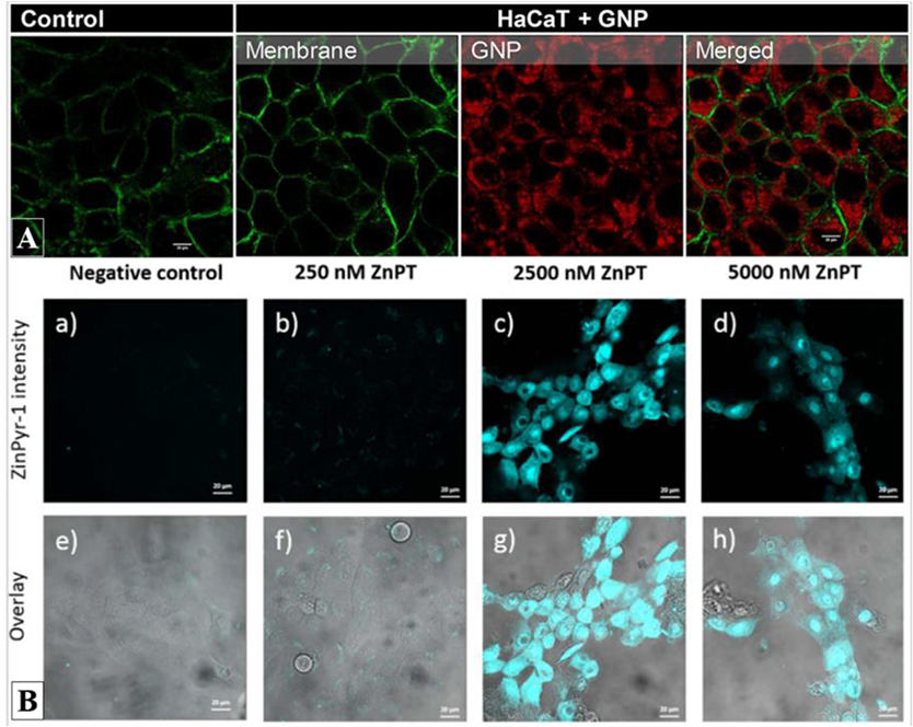
Figure 2.

Example of confocal fluorescence images of compound uptake within human keratinocytes. A) Internalization of BODIPY-labeled gold nanoparticles (red) into the cytoplasm of HaCaT keratinocytes stained with Alexa Fluor 488-conjugated with fluorescent wheat germ agglutinin (WGA) for glycocalyx membrane identification. Adapted with permission from Limon et al. Bioconjugate Chem. 29, 1060–1072 (2018) [25]. Copyright (2019) American Chemical Society; B) (top) Confocal fluorescence images of exogenous labelvZynPyr-1 (cyan) highlighting the intracellular labile zinc and its increase after application of different concentrations of zinc pyrithione (ZnPT) on HaCaT keratinocytes. (bottom) Overlay of confocal fluorescence and transmitted light images allowing to map the fluorophore’s distribution within the cells. Reprinted from Toxicol. Appl. Pharmacol. 343, Holmes et al., Imaging the penetration and distribution of zinc and zinc species after topical application of zinc pyrithione to human skin, 40-47, Copyright (2018), with permission from Elsevier [8].
