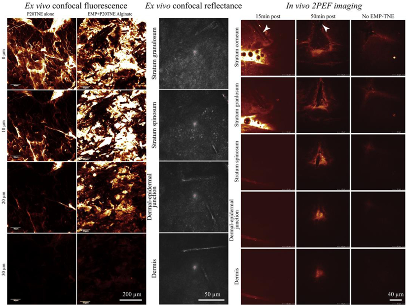
Figure 3.

Example of ex vivo confocal fluorescence, in vivo confocal reflectance and in vivo two-photon excited fluorescence images of elongated microparticles (EMP) combined with tailorable nanoemulsions (P20TNE) to alliance topical delivery of hydrophobic drug surrogates in human skin. (left) ex vivo confocal fluorescence images showing the distribution of a fluorescent lipophilic dye (DiI, incorporated within TNE) at the surface (0 μm), 10, 20 and 30 μm deep in human skin. The P20TNE alone appears to partition in the stratum corneum and furrows, whereas EMP coated with P20TNE using alginate (EMP+P20TNE Alginate) were capable of delivering detectable DiI around the dermal-epidermal junction at >30 μm deep. (middle) Ex vivo confocal reflectance images showing the presence of EMP+P20TNE Alginate through the excised living abdominal skin. (right) In vivo 2PEF images of human forearm skin after topical application TNE (alone and EMP+P20TNE Alginate) containing 6-Carboxyfluorescein (CaF) in the core droplet of the TNE. At 15 min post application, majority of TNE stayed at the surface of the skin, whereas at 50 min, more intense TNE signal released from EMP was detected at dermal-epidermal junction. Adapted from J. Control. Release, 288, Yamada et al. Using elongated microparticles to enhance tailorable nanoemulsion delivery in excised human skin and volunteers, 264-276, Copyright (2018), with permission from Elsevier [3].
