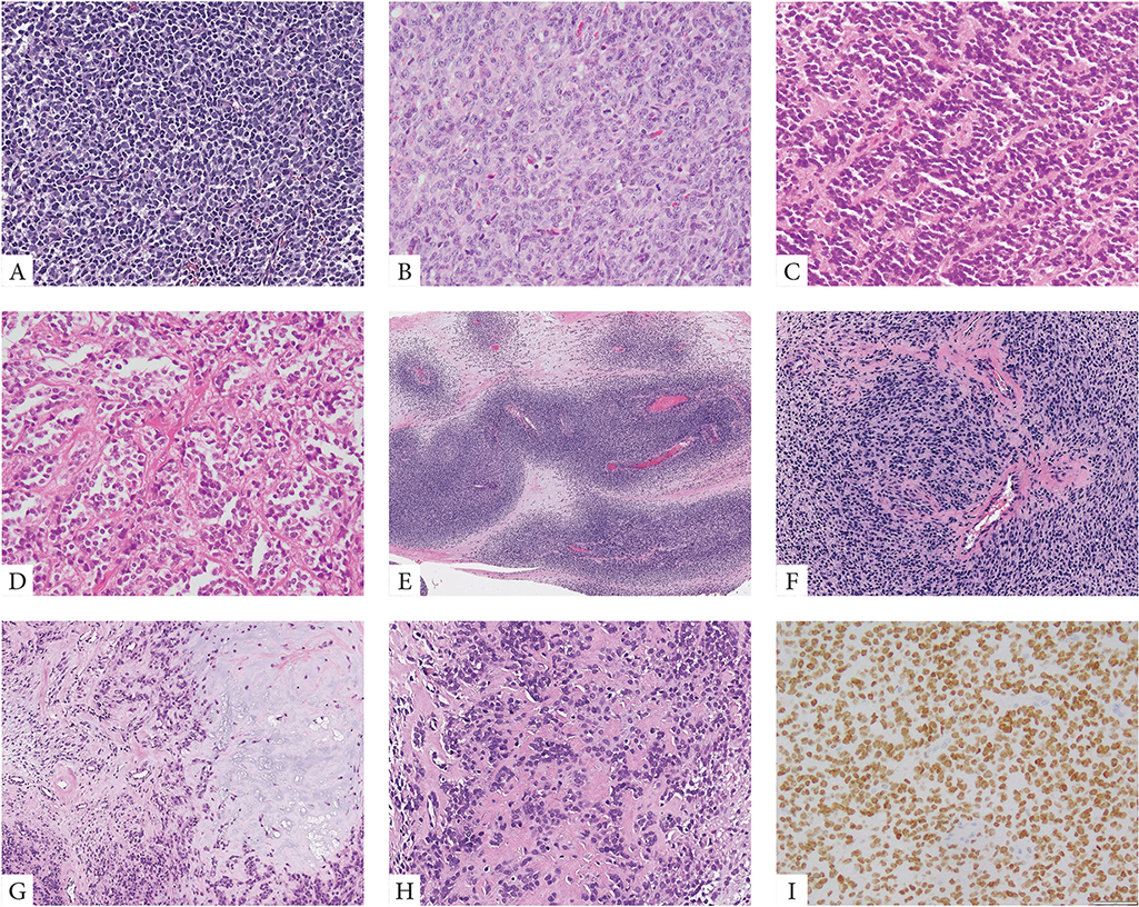
Figure 1. Morphologic spectrum of URCS with YWHAE and BCOR ITD genetic alterations.
YWHAE-NUTM2B/E positive congenital tumor composed of primitive round cells arranged in solid sheets and vague nests (A, case 4). BCOR-ITD infantile URCS showing a solid growth of undifferentiated round to ovoid cells with scant eosinophilic cytoplasm and vesicular nuclei with small nucleoli, mitotic activity is brisk (B, case 9). Alternative growth patterns included rosette formation and alveolar growth (C, D, case 12). Two cases occurred in adolescents both showing matrix formation radiographically and focally microscopically, being misinterpreted as small cell osteosarcomas (E-H, cases 19&20). The scapular bone lesion in a 16 year-old male showed a marbled appearance on low power with alternating hyper and hypocellular areas (E, case 19); at high power the primitive round cells showed a vague nesting growth and focal hyalinized stroma which in the context of SATB2 positivity was interpreted as osteoid production (F). Similarly, the paraspinal lesion showing vertebral bone involvement and marked sclerosis on radiology, was composed of primitive round cells embedded in a myxochondroid and fibrotic matrix (G, H, case 20). Strong and diffuse BCOR expression is typically seen in all cases (I).

