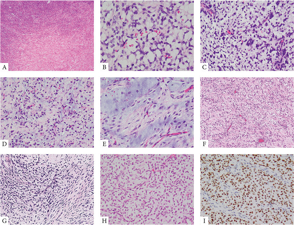
Figure 2. Morphologic spectrum of PMMTI with BCOR ITD abnormalities.
A small number of PMMTI cases showed areas of a solid round cell component limited to <30% of the mass (A, case 32). Most tumors however were diffusely myxoid with primitive round, ovoid or spindle cells floating within the extracellular matrix and associated with a delicate capillary network (B-E, cases 22,24,29). Despite low cellularity, the mitotic activity was often increased (B, case 29). Focal areas of spindling within a fibromyxoid stroma was also noted focally in some cases (F, G, cases 28,31). Focal PMMTI-like areas were noted in a subset of URCS (H, case 18). Similar to URCS, PMMTI consistently showed strong immunoreactivity for BCOR (I).

