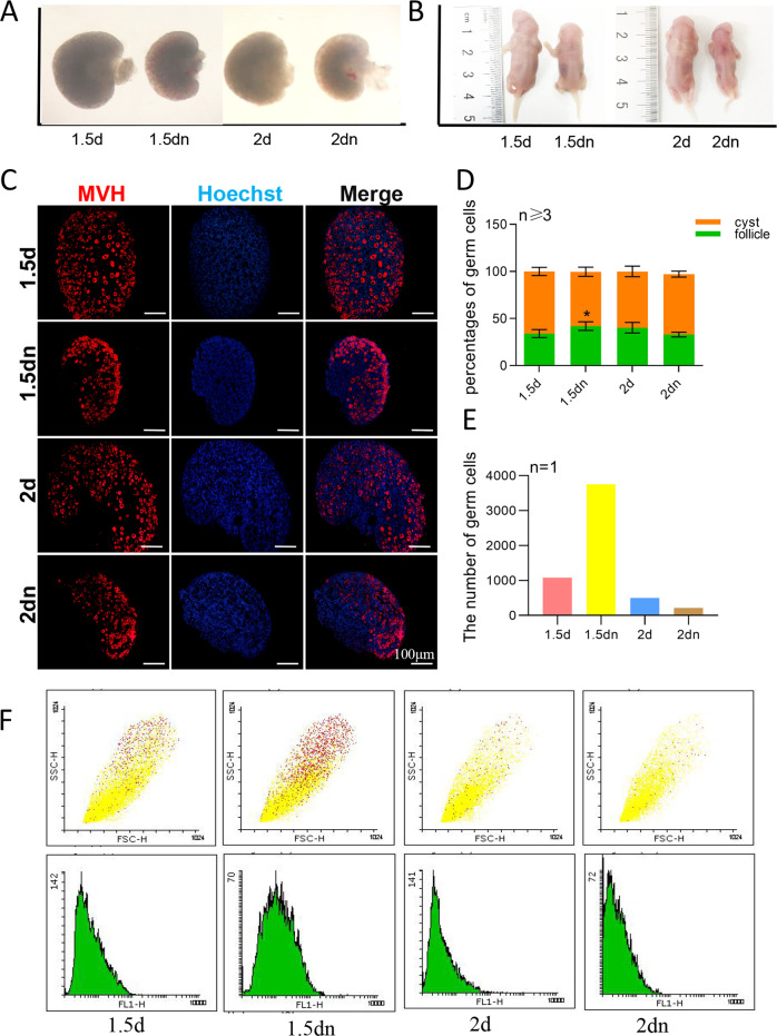
Fig. 3. Starvation induced autophagy then apoptosis in the newborn ovaries.
a Comparison of the size of the ovary after 1.5 days’ starvation and 2 days’ starvation; b Comparison of the size of the body after 1.5 days’ starvation and 2 days’ starvation; c Immunofluorescence staining of ovaries from 1.5 days’ starvation mice and 2 days’ starvation mice. Germ cells were stained red with anti-Mvh antibody, nuclei were stained blue with Hochest 33342; d Percentage of germ cells in Cyst and in primordial follicle after 1.5 days’ starvation and 2 days’ starvation; e, f Percentage of germ cells in the whole ovary after 1.5 days’ starvation and 2 days’ starvation analyzed by flow cytometry.

