
Figure 4.
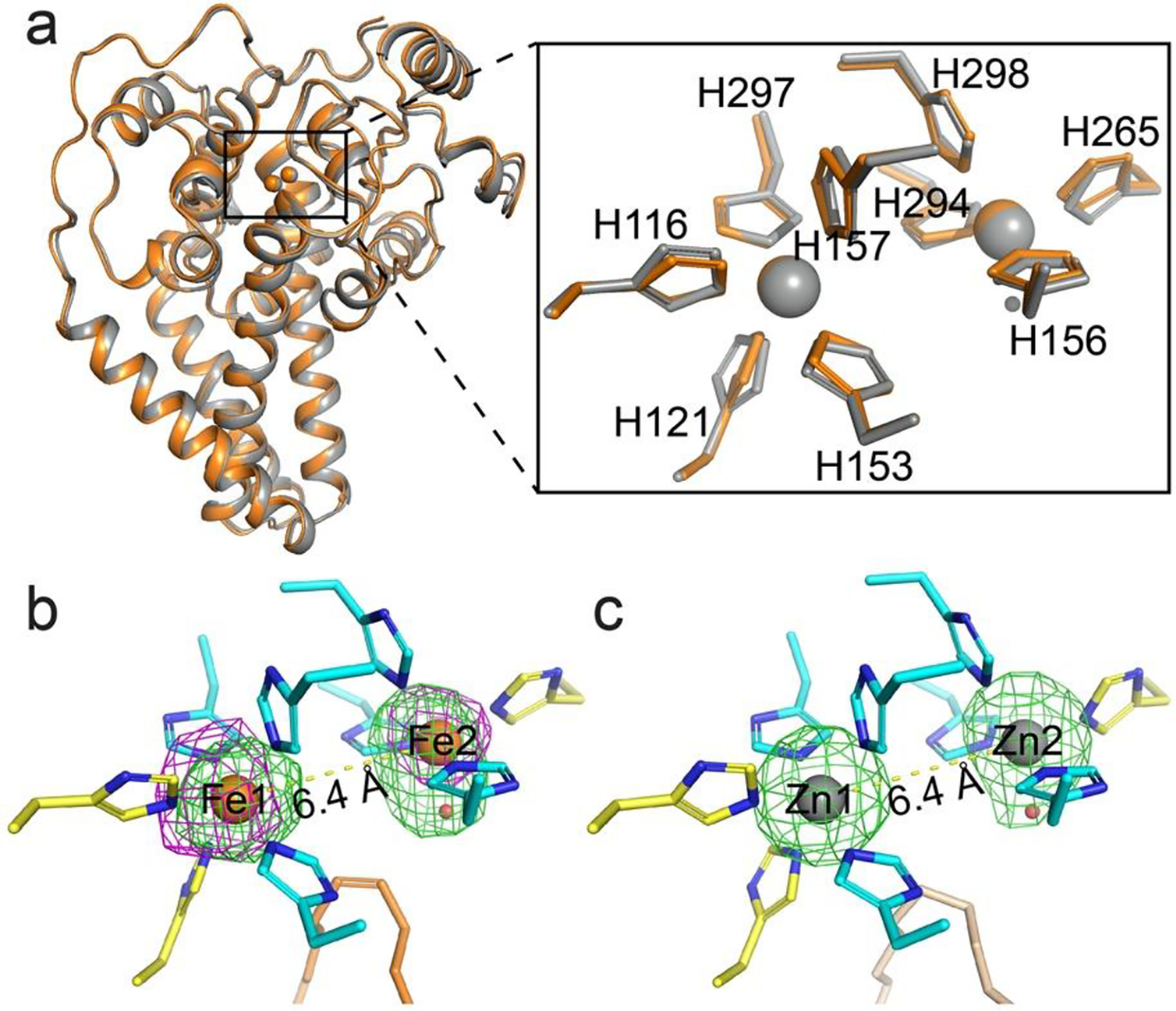
Crystal structure of iron-containing mouse SCD1. a. Structural alignment of iron-containing (orange) and zinc-containing (gray) SCD1. Inset, alignment of the dimetal center and the coordinating histidine residues. b. The reaction center is occupied by a diiron center with a unique coordination scheme that is preserved in the structure. The iron ions (orange) and a water molecule (red) are shown as spheres. Nine histidine ligands (residues from TMs in yellow; from the soluble domain in cyan) and the oleoyl-CoA (orange) are shown as sticks. Green mesh, Fo - Fc map with iron ions omitted contoured at 3 σ; magenta mesh, anomalous difference map at 3.5 σ. c. Metal coordination in the zinc-containing structure (PDB ID: 4YMK). An elongated density is also observed at Zn2. The stearoyl-CoA are shown as wheat stick. Green mesh, Fo - Fc map with zinc ions omitted contoured at 4.0 σ.