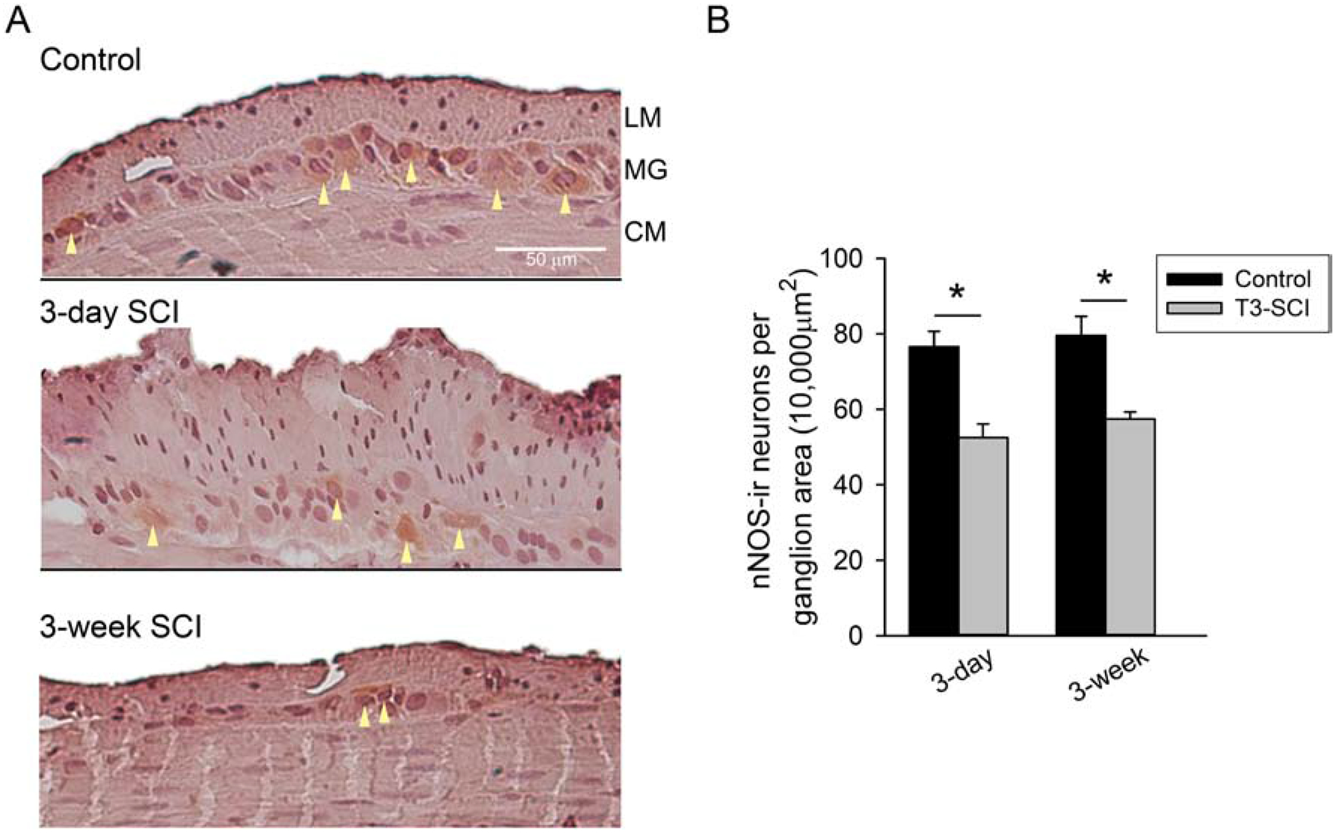
Figure 2.

A) nNOS-immunoreactive tissue sections from the distal colon of control (top), 3-day T3-SCI (middle) or 3-week T3-SCI (bottom). Representative cells exceeding staining detection threshold denoted by arrowheads. Tissues sections are from distal colon samples adjacent to those harvested from rats used for intracellular recordings (X400, scale bar = 50 μm). B) T3-SCI provokes a significant reduction in nNOS immunoreactivity in the myenteric ganglia (Values expressed as mean ± SEM, *p < 0.05 vs. post-operative time-matched controls, n = 24).
