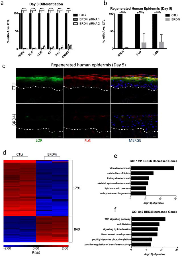
Figure 1. BRD4 is necessary for epidermal differentiation gene expression.
(a) RT-qPCR quantifying the relative mRNA expression levels of a panel of epidermal differentiation genes in scrambled control (CTLi) and BRD4 (BRD4i) siRNA treated keratinocytes after three days of differentiation. Two separate siRNAs (siRNA 1 and siRNA 2) targeting different regions of BRD4 mRNA were used (siRNA 1 n=4, siRNA 2 n=3). Statistics: t-test, ***p < 0.001. (b) RT-qPCR quantifying the relative mRNA expression levels of LOR, BRD4, and FLG in CTLi and BRD4i day 5 regenerated human epidermis. n=5 (c) Immunofluorescent staining of late differentiation markers LOR (green) and FLG (red) in CTLi and BRD4i day 5 regenerated human epidermis. Merged image includes Hoechst staining of nuclei. n=3. White Scale bar = 20μm. (d) Heatmap generated for replicate (n=2) RPKM normalized RNASequencing data from CTLi and BRD4i keratinocytes differentiated for 3 days. The expression of genes significantly increased (red) or decreased (blue) is shown. Differential expression was determined with FDR ≤ 0.05 and fold change ≥ 2 vs. CTLi. Graphs are displayed in log2 scale. (e) Gene ontology (GO) term enrichment for the 1,791 genes significantly decreased in BRD4 knockdown cells. (f) Gene ontology (GO) term enrichment for the 840 genes significantly increased in expression in BRD4 knockdown cells.

