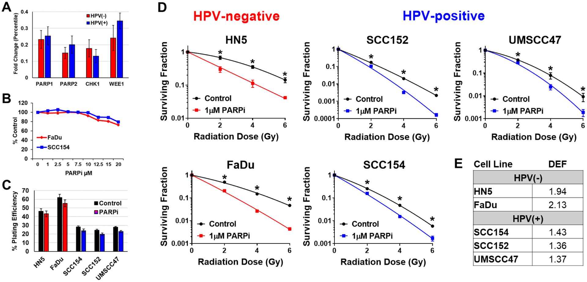
Figure 1: PARPi radiosensitizes HNSCC cells with little cytotoxicity as a single agent.

(A) In vivo shRNA screening of tumors generated from five different HNSCC cell lines (2 HPV(+) and 3 HPV(−)) comparing the difference in magnitude of radiosensitization between HPV(−) and HPV(+) tumor models following the expression of the shRNA constructs for PARP1, PARP2, Chk1 or Wee1. (B) MTS assay at 24h after treatment with niraparib (PARPi) at doses of 0–20μM with 6 replicates/dose. (C) Plating efficiencies in HPV(−) (red) and HPV(+) (blue) cell lines taken from CSAs shown in D. (D) Cells were treated with 1μM niraparib (PARPi) for 1h then irradiated with 0–6Gy and replated 24h later for colony formation. (E) Radiation dose enhancement factors (DEF) calculated at surviving fraction of 0.5 in HPV(−) and HPV(+) cell lines following treatment with niraparib (PARPi) and radiation.
