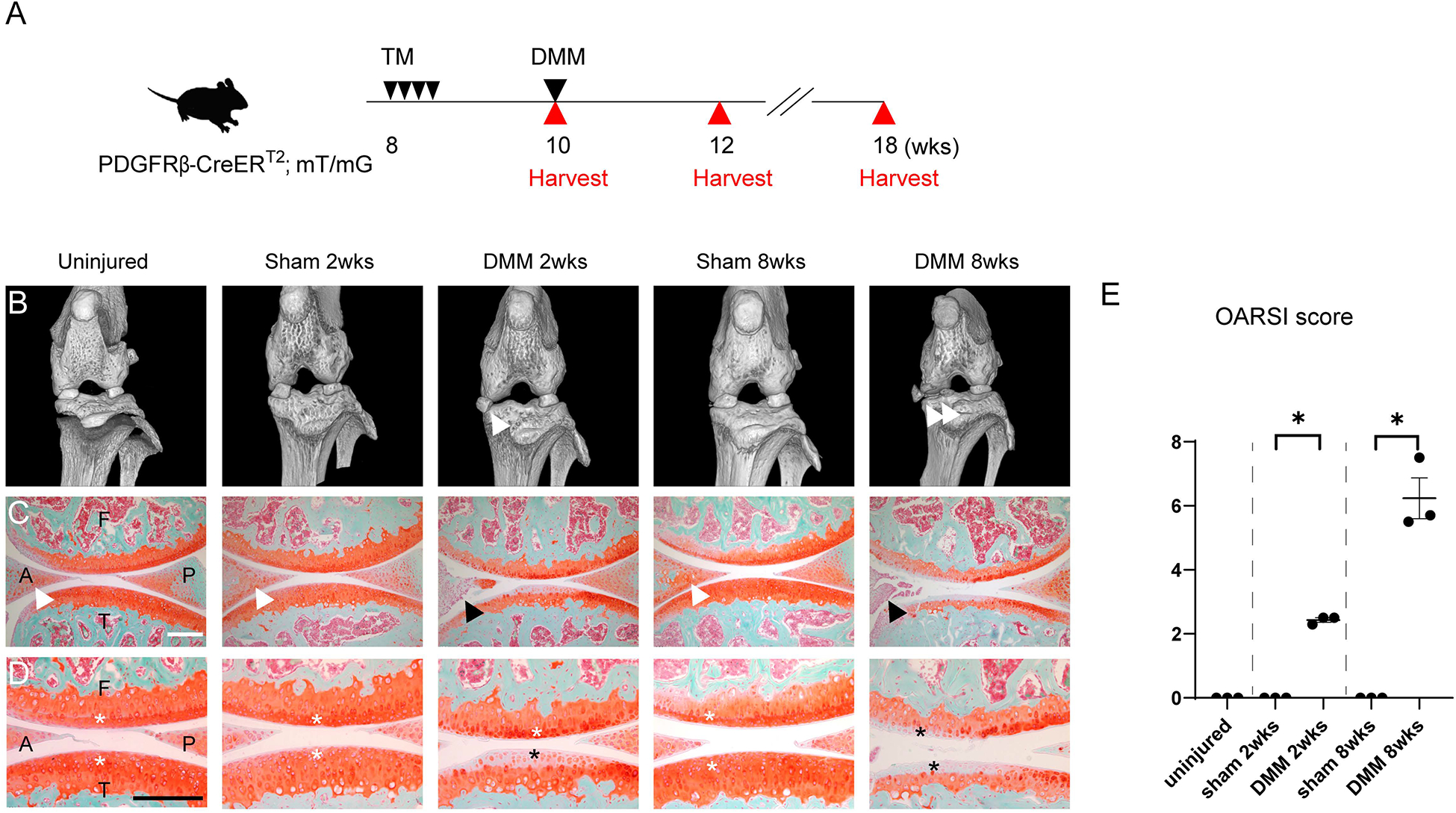
Figure 1.

Validation of DMM induced osteoarthritis within PDGFRβ-CreERT2 reporter animals. (A) Schematic of experiments, including tamoxifen (TM) injection daily to PDGFRβ-CreERT2; mT/mG mice for 4 consecutive days at 8 weeks of age, followed by DMM surgery at 10 weeks of age. Analyses were performed at 2 and 8 weeks after DMM. (B) Micro computed tomography (microCT) images of the left knee joints of PDGFRβ-CreERT2; mT/mG mice. White arrowheads indicate the displaced medial meniscus at 2 and 8 weeks post-operative among DMM treated groups. (C) Sagittal sections of the left knee joints of PDGFRβ-CreERT2; mT/mG mice at the level of medial compartments. Safranin O- fast green staining. Anterior horns of the medial meniscus are indicated in the uninjured, sham 2 weeks and sham 8 weeks group with white arrowheads and in DMM 2 and 8 weeks with black arrowheads. (D) High magnifications of Safranin O- fast green staining. Cartilage surfaces were marked with white asterisks in control groups, with black asterisks in DMM groups. (E) OARSI scores. A: anterior horn of the medial meniscus, P: posterior horn of the medial meniscus, F: femur, T: tibia. Scale bars: 100μm. N=3 mice per group. *p<0.05.
