Figure 2.

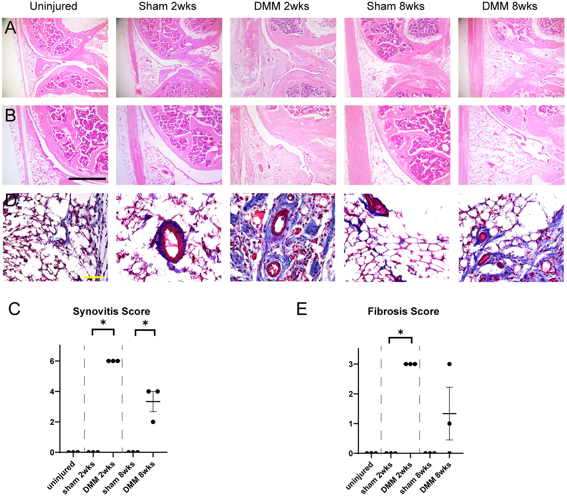
Histologic changes within the IFP after DMM within PDGFRβ-CreERT2 reporter animals. (A,B) Sagittal sections of the left stifle joints of PDGFRβ-CreERT2; mT/mG animals at the level of posterior cruciate ligaments. Hematoxylin and eosin (H&E) staining at low (A) and (B) high magnifications. Scale bars: 200 μm. (C) Synovitis scores within PDGFRβ-CreERT2; mT/mG mice. (D) Masson Trichrome staining of the infrapatellar fat pad (IFP). Scale bars: 50 μm. (E) Fibrosis scores o within PDGFRβ-CreERT2; mT/mG mice. N=3 mice per group. *p<0.05.
