
Figure 3.
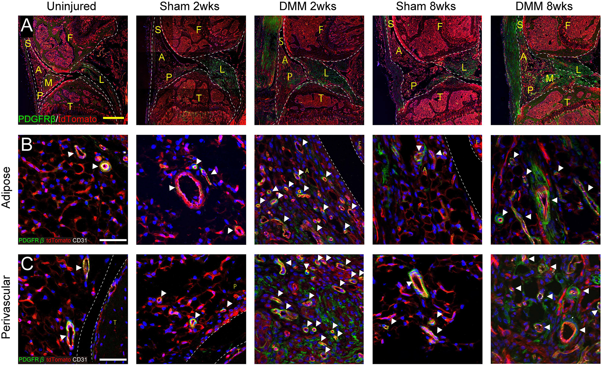
PDGFRβ reporter activity with or without DMM. (A) Immunofluorescent images of the knee joints among PDGFRβ-CreERT2 reporter mice at the level of posterior cruciate ligaments. PDGFRβ reporter activity appears green, while all other cells appear red. DAPI nuclear counterstain appears blue. Scale bars: 200 μm. (B,C) High magnifications of PDGFRβ-CreERT2 reporter activity with CD31 immunofluorescent staining, within either (B) adipose or (C) perivascular areas. PDGFRβ reporter activity appears green, while CD31 immunostaining appears white. White arrowheads indicate vessels. Scale bars: 50 μm. S: Synovium, A: adipose area, P: perivascular area, F: femur, T: tibia, L: posterior cruciate ligament, M: meniscus. N=3 mice per group.