
Figure 5.
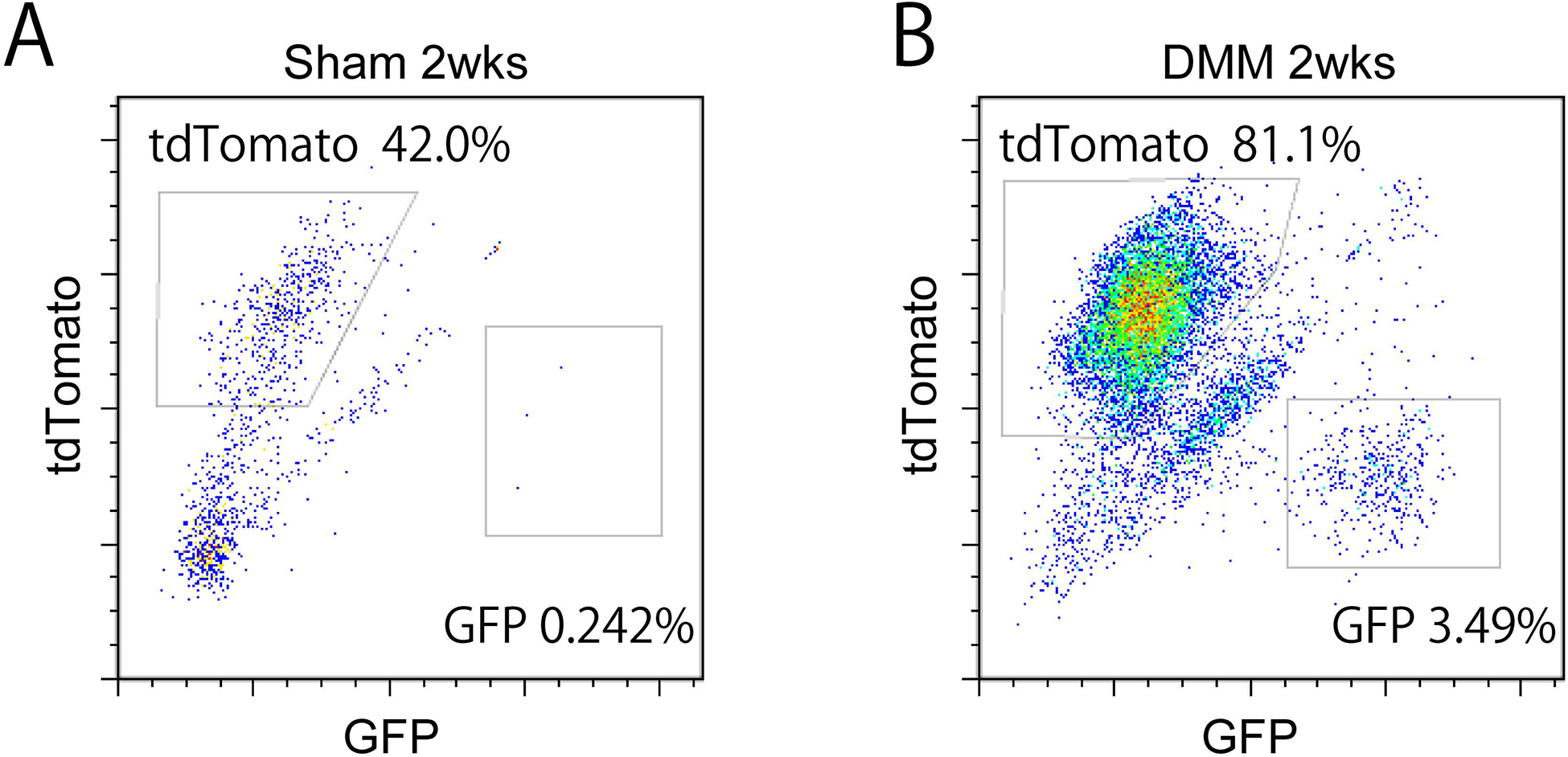
Flow cytometry analysis and quantification of PDGFRβ reporter among microdissected IFP. Samples analyzed at 2 weeks post sham or DMM surgery among PDGFRβ reporter animals. Frequency of mGFP+ and TdTomato+ cells was assessed. (A) 2 weeks post sham surgery, and (B) post DMM surgery. N=11 pooled mice per group.