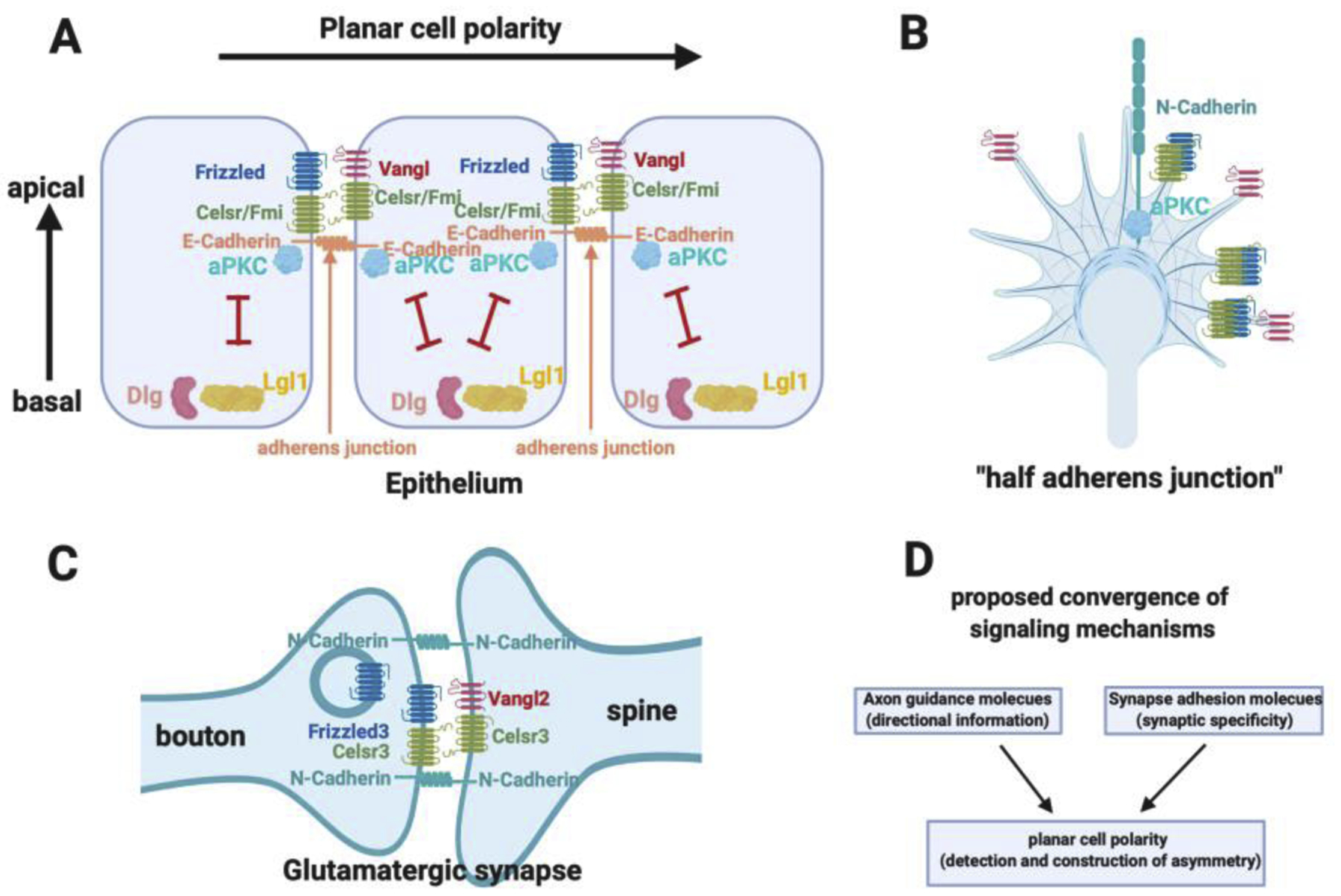
Figure 4. Planar cell polarity signaling in growth cone guidance and synapse formation.

A. Schematics of planar cell polarity and apical-basal polarity and their interactions. E-Cadherin is located in the adherens junctions where aPKC is localized and activated. Planar cell polarity complexes are built at the adherens junctions.
B. The growth cone has the same planar cell polarity and apical basal polarity components and similar molecular organizations as the adherens junctions. The growth cone has N-Cadherin.
C. The glutamatergic synapses have the same planar cell polarity and apical basal polarity components and similar molecular organization as the adherens junctions.
D. The hypothesis that cell polarity pathways may be a common mechanism mediating the function of axon guidance molecules and synapse adhesion molecules, which provide directional and positional information to provide specificity for brain wiring.
