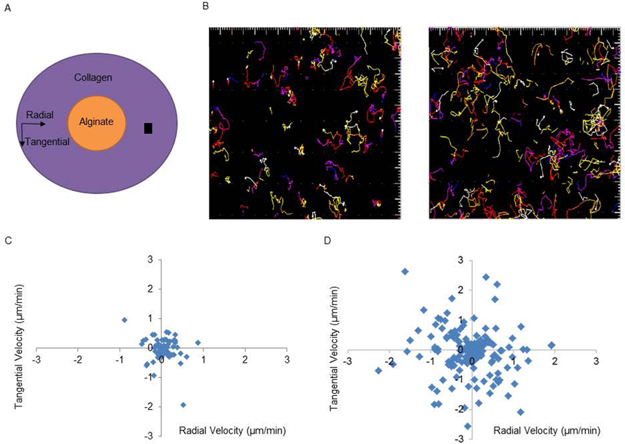
Figure 3. GM-CSF mediated chemokinesis of bone marrow derived dendritic cells in vitro.

(A) Alginate gels with or without GM-CSF were placed in a petri dish and surrounded with collagen containing labeled murine bone marrow derived dendritic cells. The cartoon depicts a transverse section of the petri dish with the purple color representing the collagen and DC while the orange color denotes the alginate gel and the black square represents an imaging window as seen in (B). The imaging window was randomly selected. (B) Individual paths of cells in a representative experiment exposed to control (no GM-CSF) or GM-CSF containing alginate hydrogels viewed at 20x. The average velocity of the cells was calculated from initial and final position values and is plotted for control gels (C) and GM-CSF releasing gels (D). Chemotaxis toward the alginate is given as the positive radial coordinate. Each dot reflects the velocity of 1 cell and each plot is representative of three experiments.
