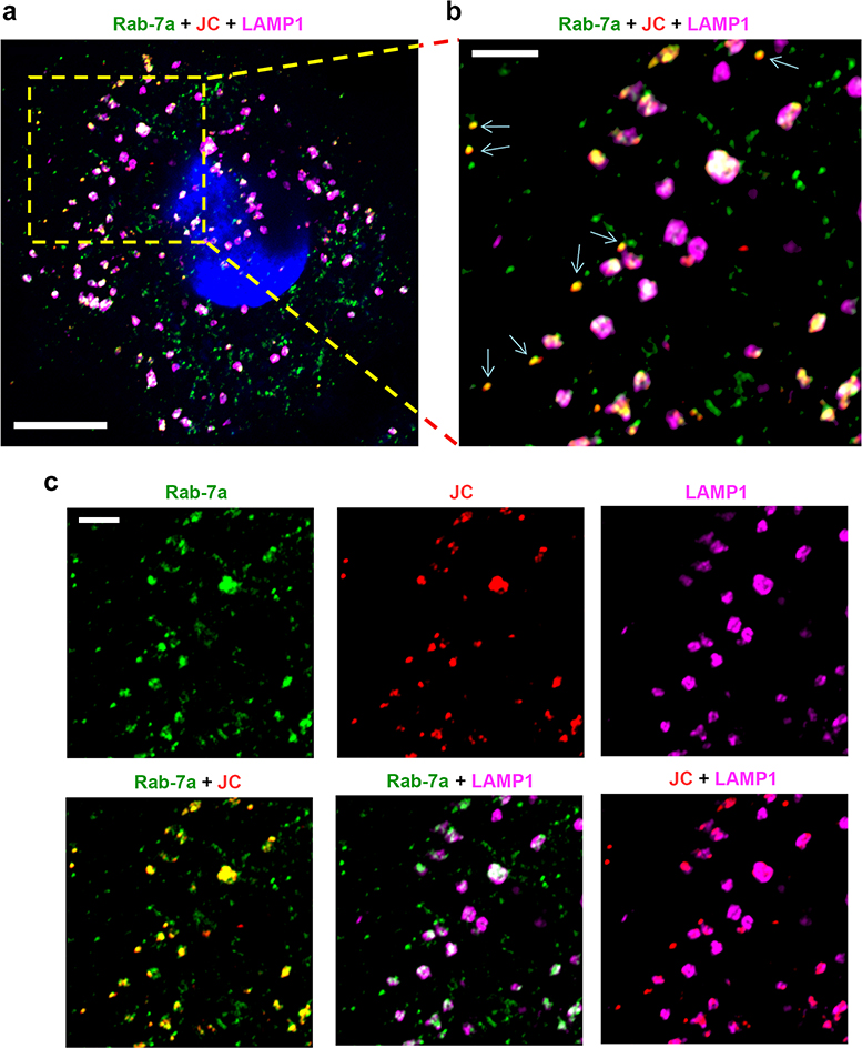
Extended Data Fig. 7.
Endolysosomal location of HAV capsids in UGCG-KO1.3 cells.
a. Airyscan microscopic image of 18f-infected UGCG-KO1.3 cells labelled with antibodies to Rab-7a (green), HAV (JC, red) and LAMP1 (magenta). Virus was adsorbed to cells for 2 hrs at 37 ºC, which were then washed with PBS and reincubated at 37 ºC for 4 hrs prior to fixation. Scale bar = 10 μm. b. Expanded view of the area bordered by the yellow rectangle in panel (a) demonstrating that most HAV capsids are localized within endolysosomes staining for Rab-7a+ and/or LAMP1+. The arrows indicate capsids present in late endosomes staining only for Rab-7a. Scale bar = 3 μm. c. Single and dual fluorescence signals present in the image shown in panel (b). Scale bar = 3 μm. Images are representative of findings in two independent experiments with similar results.

