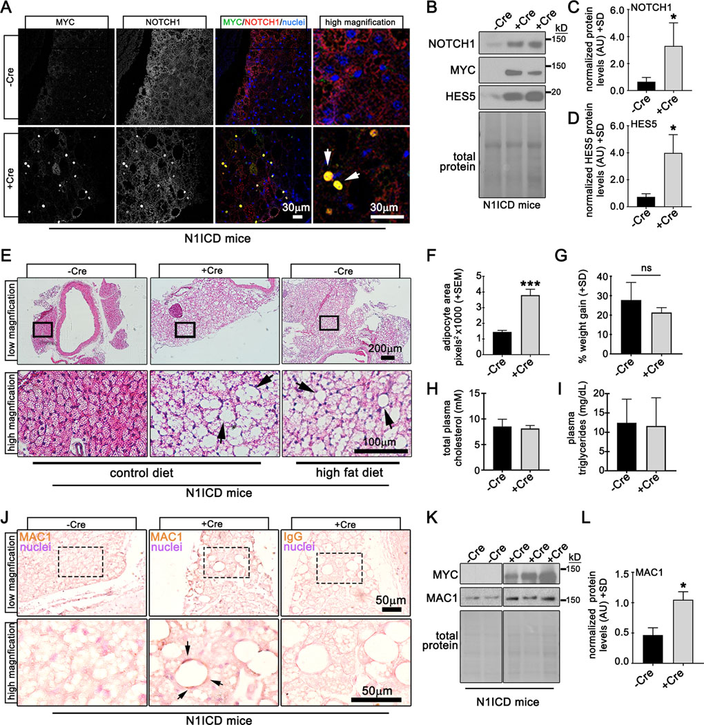
Figure 4. Notch1 activation promotes pathological conversion of tPVAT in mice fed a control diet.

A) Verification of myc-tagged N1ICD transgene was performed by confocal immunofluorescence of tPVAT from N1ICD (-Cre) or Adipoq-Cre;N1ICD (+Cre) mice for indicated proteins. Arrows show nuclear colocalization of the myc epitope and N1ICD. B) Immunoblot of tPVAT from -Cre or +Cre N1ICD mice, with quantification of protein levels (C-D). E) H&E staining of aorta with tPVAT from -Cre or +Cre mice on the indicated diets (arrows, lipid accumulation). F) Quantification of adipocyte area from iWAT from mice fed a control diet for 12 weeks, indicating adipocyte hypertrophy. G) Weight gain for each group was not different. Total plasma cholesterol (H) and triglycerides (I) from -Cre or +Cre N1ICD mice are shown. J) PVAT sections were immunostained to detect macrophages (MAC1), which were prominent in +Cre mice compared to -Cre controls. Negative control with IgG only is shown. K-L) Immunoblot to detect MAC1 in PVAT verifies an increase in +Cre N1ICD mice. Graphed are means +SD. Data were analyzed using Student’s t-test with post-hoc Tukey’s range test. *P≤0.05 ***P≤0.001, ns = not significant
