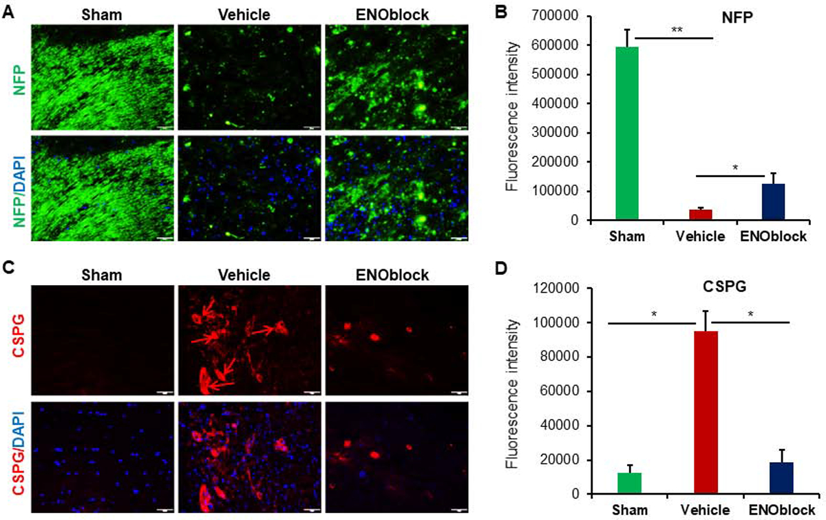
Fig. 4.

ENOblock treatment induces neuroprotection in SCI. (A) Neurofilament protein (NFP, green) and DAPI (blue) nuclear staining of SCI tissue at the injury site. Panel shows representative images taken at 20× magnification. (B) Mean fluorescence intensity from three representative areas shows significantly decreased NFP expression in vehicle treated tissue. There is also a significant increase in NFP expression indicating the recovery with ENOblock treatment as compared to vehicle treated controls (paired t-test, two-tailed, *p < 0.05, **p<0.01; n=3–5). (C) Chondroitin sulfate proteoglycan (CSPG, red; arrows) and DAPI (blue) nuclear staining of SCI tissue at the injury site. Representative images were taken at 20× magnification. (D) Mean fluorescence intensity shows a significant increase in CSPG in vehicle treated rats that is significantly reduced with ENOblock treatment (paired t-test, two-tailed, *p < 0.05; n=4–5).
