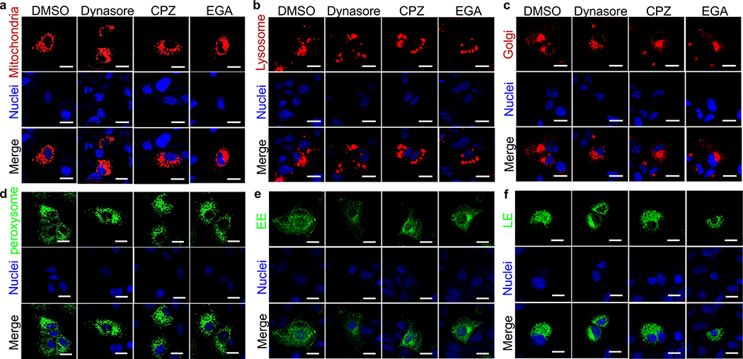
Extended Data Fig. 10.
Effect of endocytosis inhibitors in different cellular compartments.
Huh7 cells in 8-wells chamber slides were infected with the CellLight Bacman 2.0 reagents fused to GFP (green) or RFP (red) at a multiplicity of infection of 30 particles per cell, incubated at 37°C for 10–12 h, treated with 80 mM Dynasore hydrate, 5mg/ml Chlorpromazine hydrochloride solution, 10 mM EGA, or a similar volume of DMSO vehicle as negative control, and incubated for additional 12–14 h at 37°C. Nuclei were stained with DRAQ5 (blue), cells were fixed with 4% PFA, coverslips mounted with ProLong Gold antifade reagent, and slides analyzed in a LSM 700 confocal microscope. Micrographs were taken using a 63X oil objective. Cells were infected with CellLight Bacmam 2.0 driving the expression of markers of: a, Mitochondria (leader sequence of E1 alpha pyruvate dehydrogenase fused to RFP); b, Lysosomes (Lamp1 fused to RFP); c, Golgi (human Golgi-resident enzyme N-acetylgalactosaminyltransferase 2 fused to RFP); d, Peroxisomes (peroxisomal C-terminal targeting sequence fused to GFP); e, Early endosomes (EE, Rab5a fused to GFP); or f, Late Endosomes (LE, Rab 7a fused to GFP). Scale Bars represent 25μm. Results are representative of 3 independent experiments.

