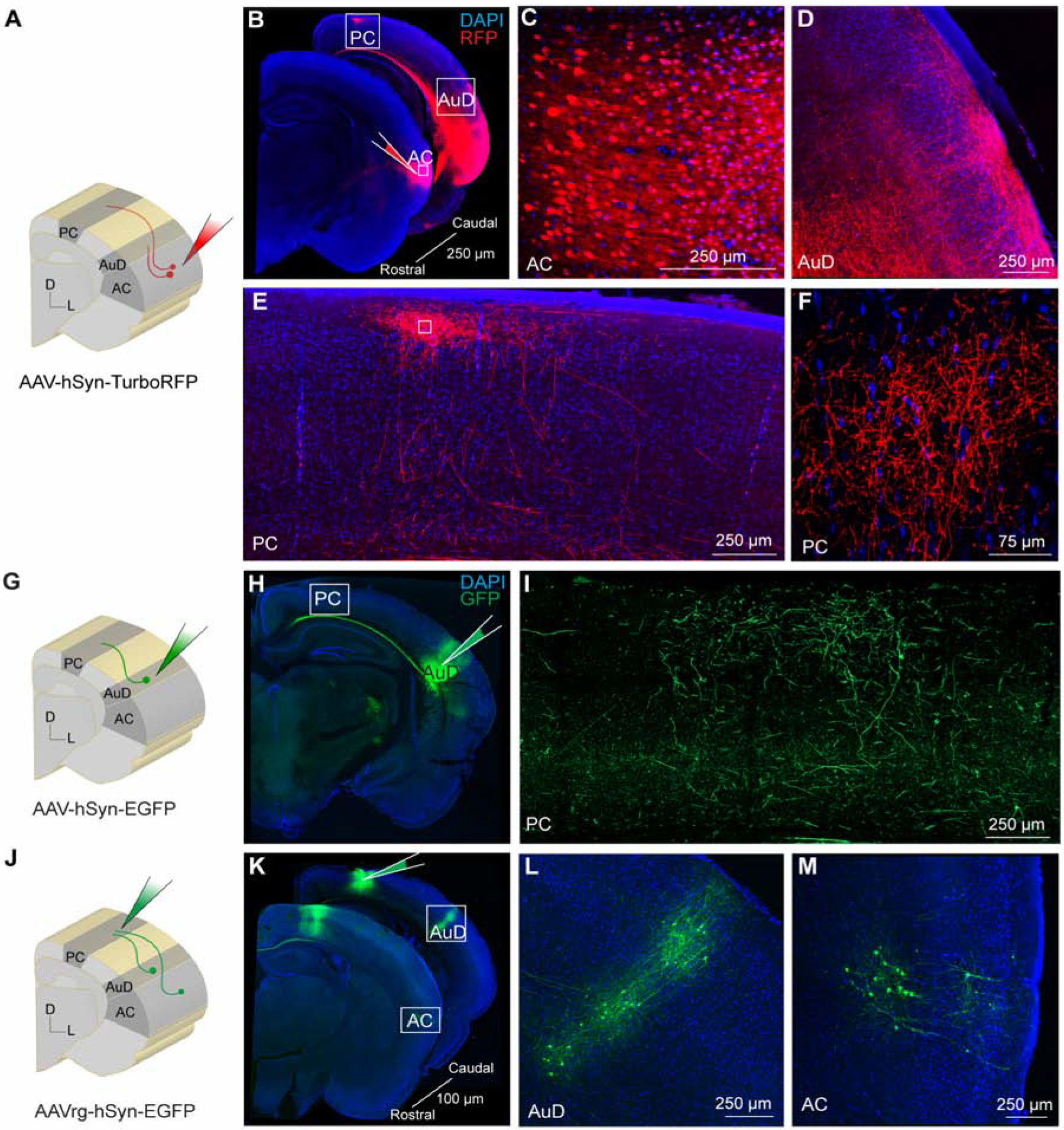
Figure 2. Auditory cortical neurons project to the parietal cortex.

A) Schematic diagram showing auditory cortex site of anterograde AAV-hSyn-TurboRFP injection. B) Brain slices showing the injection site in auditory cortex and anterograde labeling in secondary dorsal auditory area and parietal cortex. C) Expanded inset of auditory cortex showing high-magnification of labeled cell bodies within the injection site. D) Expanded inset of secondary dorsal auditory area showing dense axonal labeling. E) Expanded inset of parietal cortex showing dense axonal labeling. F) Expanded inset of parietal cortex at higher magnification. G) Schematic diagram showing secondary dorsal auditory area site of anterograde AAV-hSyn-EGFP injection. H) Brain slice showing the injection site in secondary dorsal auditory area. I) Expanded inset showing high-magnification of axonal labeling in parietal cortex. J) Schematic diagram showing parietal cortex site of retrograde AAV-hSyn-EGFP injection. K) Brain slices showing the injection site in parietal cortex and retrograde labeling in secondary dorsal auditory area and auditory cortex. L) Expanded inset of secondary dorsal auditory area showing cell body labeling. M) Expanded inset of auditory cortex showing cell body labeling. Core auditory cortex (AC), secondary dorsal auditory area (AuD), parietal cortex (PC).
