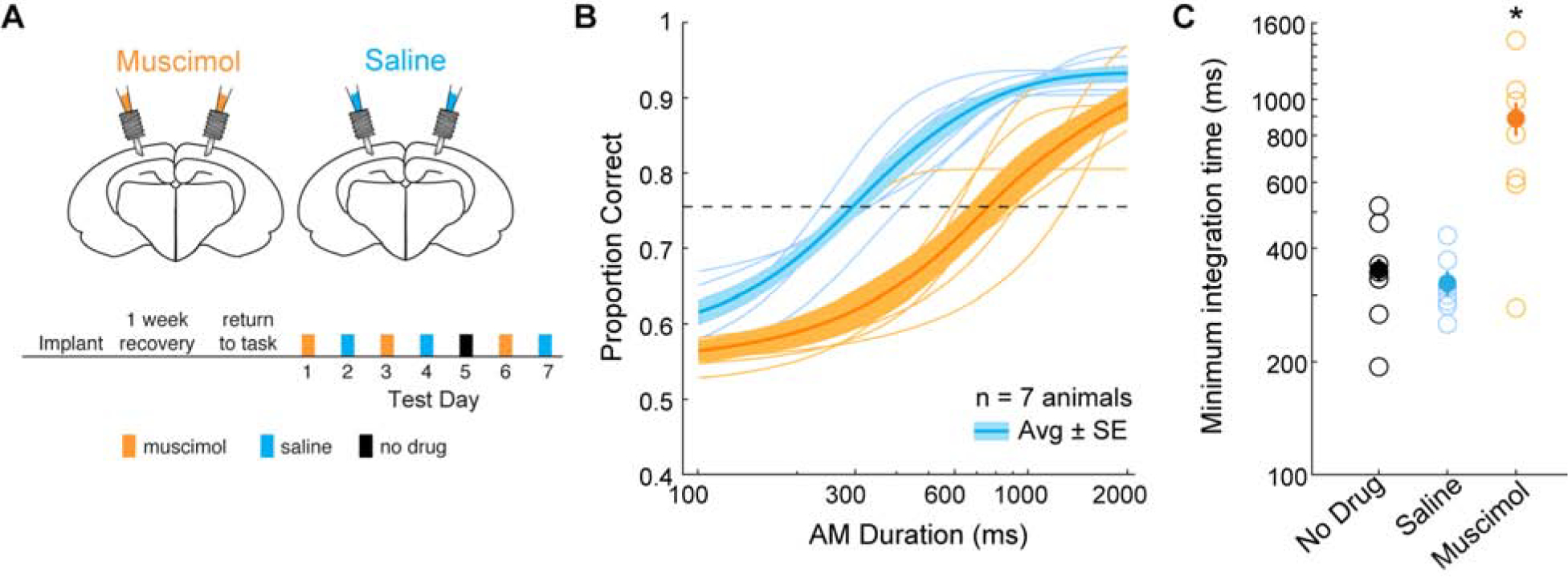
Figure 3. Inactivation of parietal cortex increases auditory integration time.

A) Schematic of cannula implant over parietal cortex and timeline of experimental test sessions. Infusions of muscimol and saline into parietal cortex alternated across test session days. Prior to the last muscimol test session, neither muscimol or saline were infused. B) Average psychometric functions across all animals (thick lines) and average psychometric functions from each animal during muscimol (orange) and saline (blue) infusion sessions (thin lines). The shaded regions represent average ± SE. See text for statistical comparisons. C) Distribution of calculated minimum integration times from each animal as a function of infusion condition. Post-hoc analyses revealed minimum integration times under muscimol (orange) were significantly different from no drug (black) (two-tailed t-test; Holm-Bonferroni-corrected; p = 0.02, t = 3.02) and saline (blue) (two-tailed t-test; Holm-Bonferroni-corrected; p = 0.01, t = 3.48) sessions. See also Figure S2.
