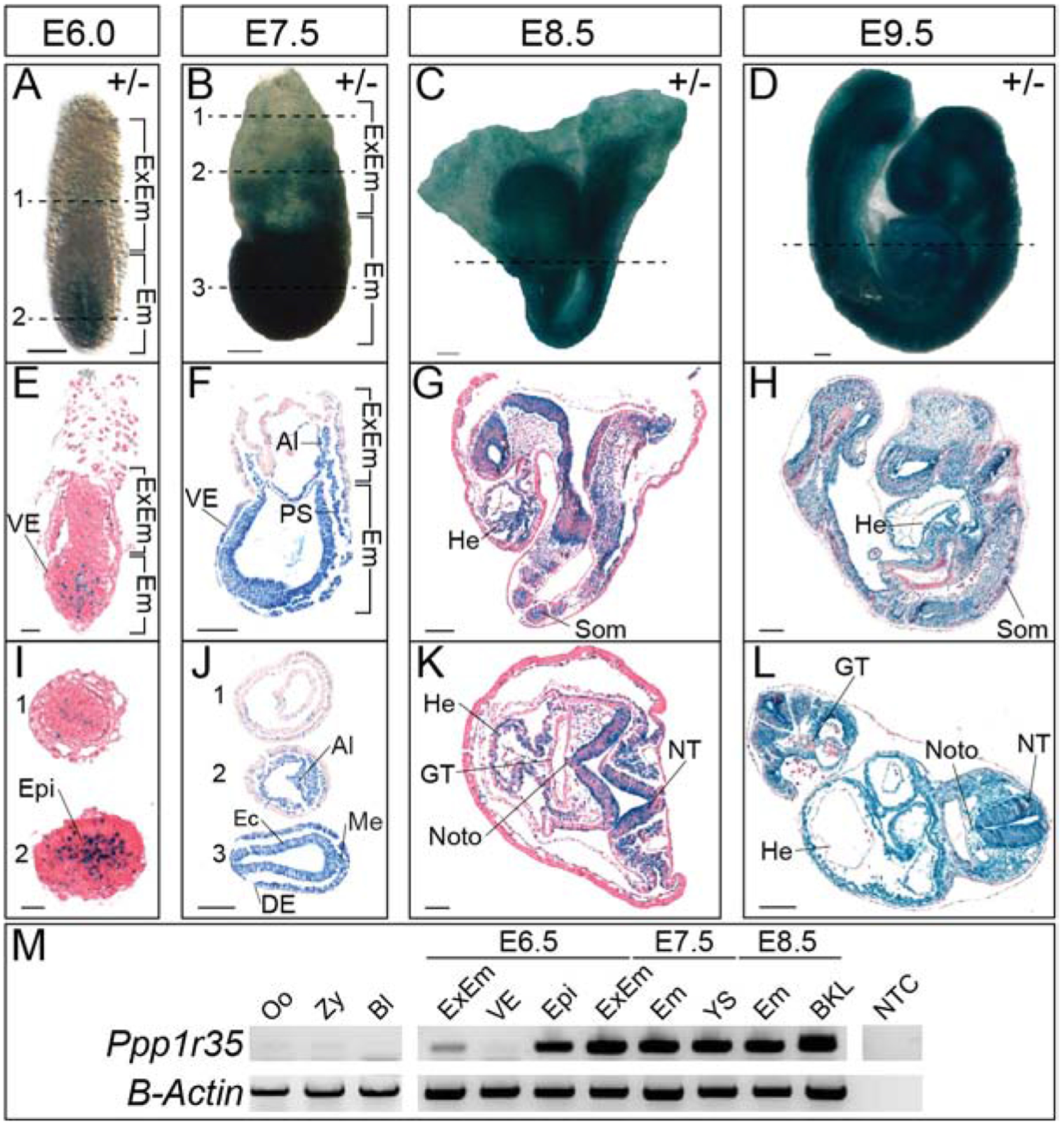
Figure 1: Ppp1r35 expression during embryonic development.

X-gal stained heterozygous embryos at E6.0 (A), E7.5 (B), E8.5 (C), and E9.5 (D). Sagittal (E-H) or transverse (I-L) of X-gal stained heterozygotes at E6.0–9.5. I) At E6.0, 1 and 2 denote sections indicated by the dashed line in (A). J) At E7.5, 1–3 denote sections indicated by the dashed lines in (B). M) RT-PCR of Ppp1r35 in various tissues and at the developmental stages denoted. β-actin is a loading control. +/− denotes Ppp1r35 heterozygous embryos. ExEm; extra-embryonic, Em; embryonic, VE; visceral endoderm, Epi; epiblast, Al; allantois, PS; primitive streak, Ec; ectoderm, DE; definitive endoderm, Me; mesoderm, He; heart, Som; somites, GT; gut tube, Noto; notochord, NT; neural tube, Oo; oocyte, Zy; zygote, Bl; blastocyst, YS; yolk sac, BKL; brain-kidney-liver, NTC; no template control. Scale bars in A-D, F-H, J, and L = 100μm, E&I = 20μm, and K= 50μm.
