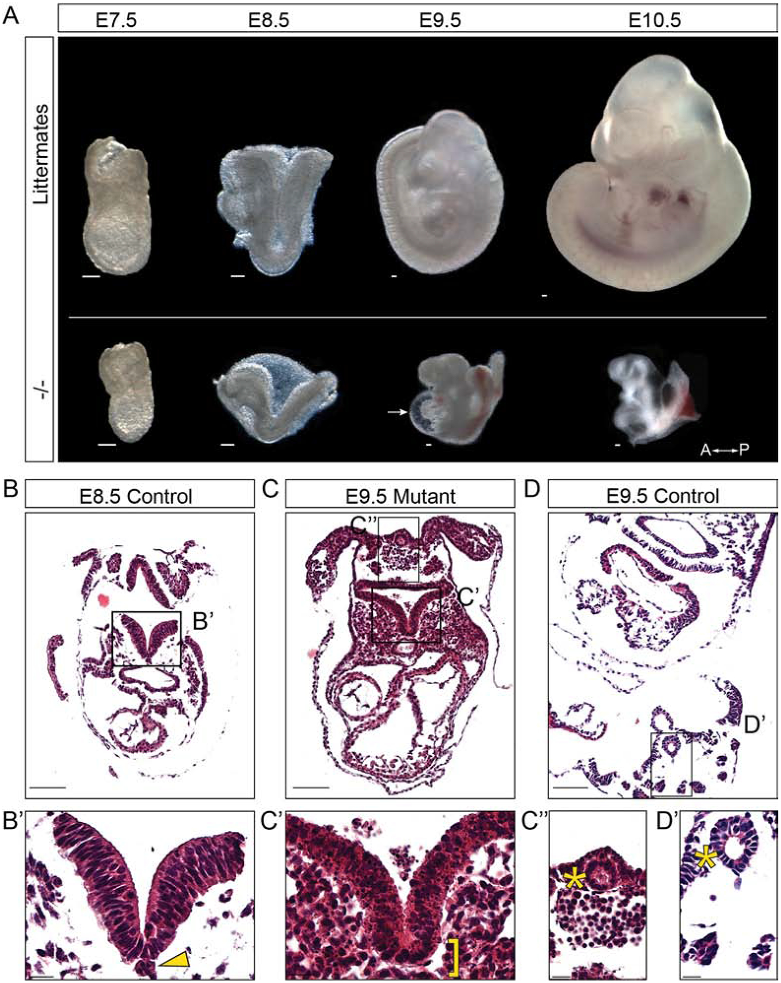
Figure 2: Ppp1r35 knockout mouse embryos show severe morphological defects compared to their littermates.

A) Ppp1r35 mutant embryos compared to littermates collected at E7.5–10.5. Hematoxylin & eosin staining performed on transverse sections of E8.5 (B,B’) and E9.5 (D,D’) control embryos compared to an E9.5 mutant (C,C’,C”) embryo. Arrow denotes the pericardial sac. A-P indicates anterior and posterior orientation of the embryos. The arrowhead points to notochord and bracket indicates expected location of the notochord. The asterisk indicates the closed hindgut of the E9.5 mutant and E9.5 control. −/− denotes Ppp1r35 homozygous embryos. Littermates or ctrl denotes embryos wild type or heterozygous for Ppp1r35. Scale bars in A,B,C,D = 100μm and B’,C’,C”,D’ = 20μm.
