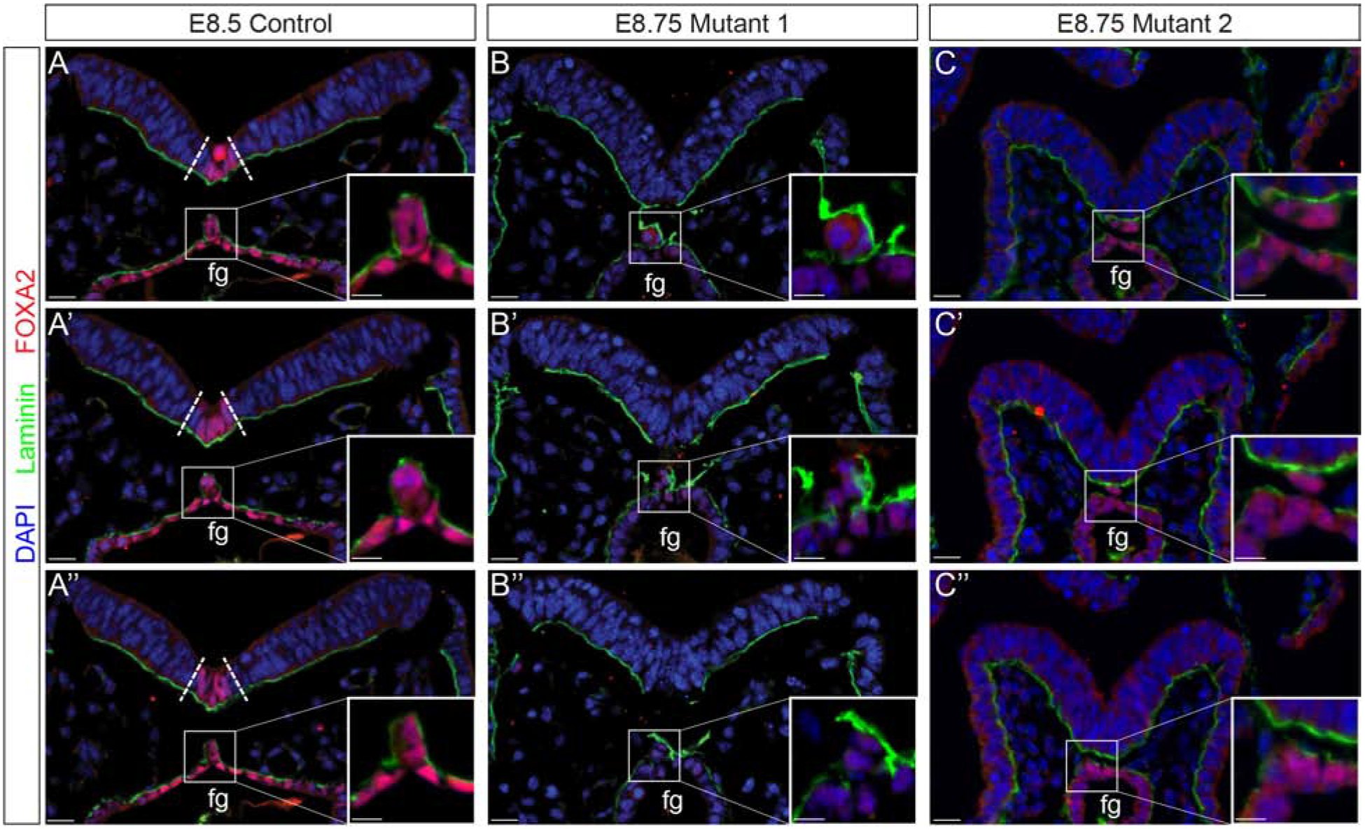
Figure 5: Notochord morphogenesis is disrupted in Ppp1r35 mutant embryos.

Immunofluorescent analysis of laminin (green), FOXA2 (red), and nuclei counterstained with DAPI (blue). Consecutive sections through a stage-matched E8.5 control reveals a FOXA2-positive floor plate (boundary defined by dotted lines) and a contiguous notochord that has laminin distributed laterally and dorsally (A-A”). Although the two E8.75 two mutants vary slightly they both lack a FOXA2-positive floor plate and display altered notochord resolution (B-B”, C-C”). Fg; foregut. Ctrl denotes embryos wild type or heterozygous for Ppp1r35. −/− denotes Ppp1r35 homozygous embryos. Boxes indicate a higher magnification of the notochord. FOXA2-labeled mutant embryos: n=3. Scale bars = 20 μm.
