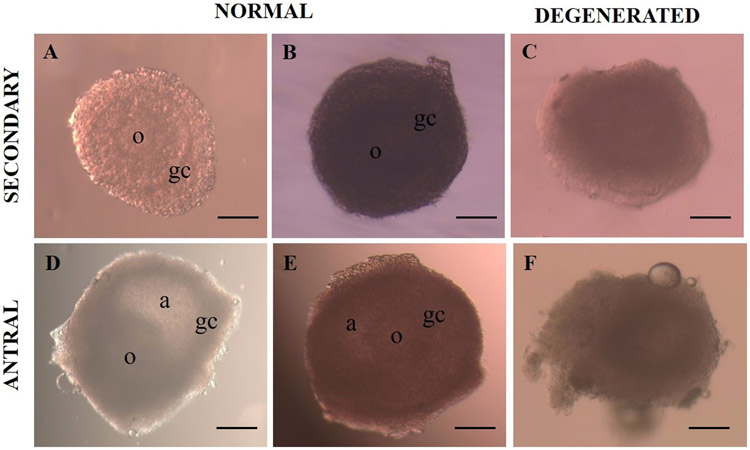
Fig. 1.
Representative images of secondary and early antral follicles. Morphologically normal, translucent follicles, with intact membrane and bright and homogenous surrounding granulosa cells (A, B, D and E). Degenerated follicles, with irregular shape, dark oocyte and granulosa cells, integrity loss and diameter reduction (C and E). a: antrum, o: oocyte, gc: granulosa cells (original magnification 10×). Scale bar = 200 μm.

