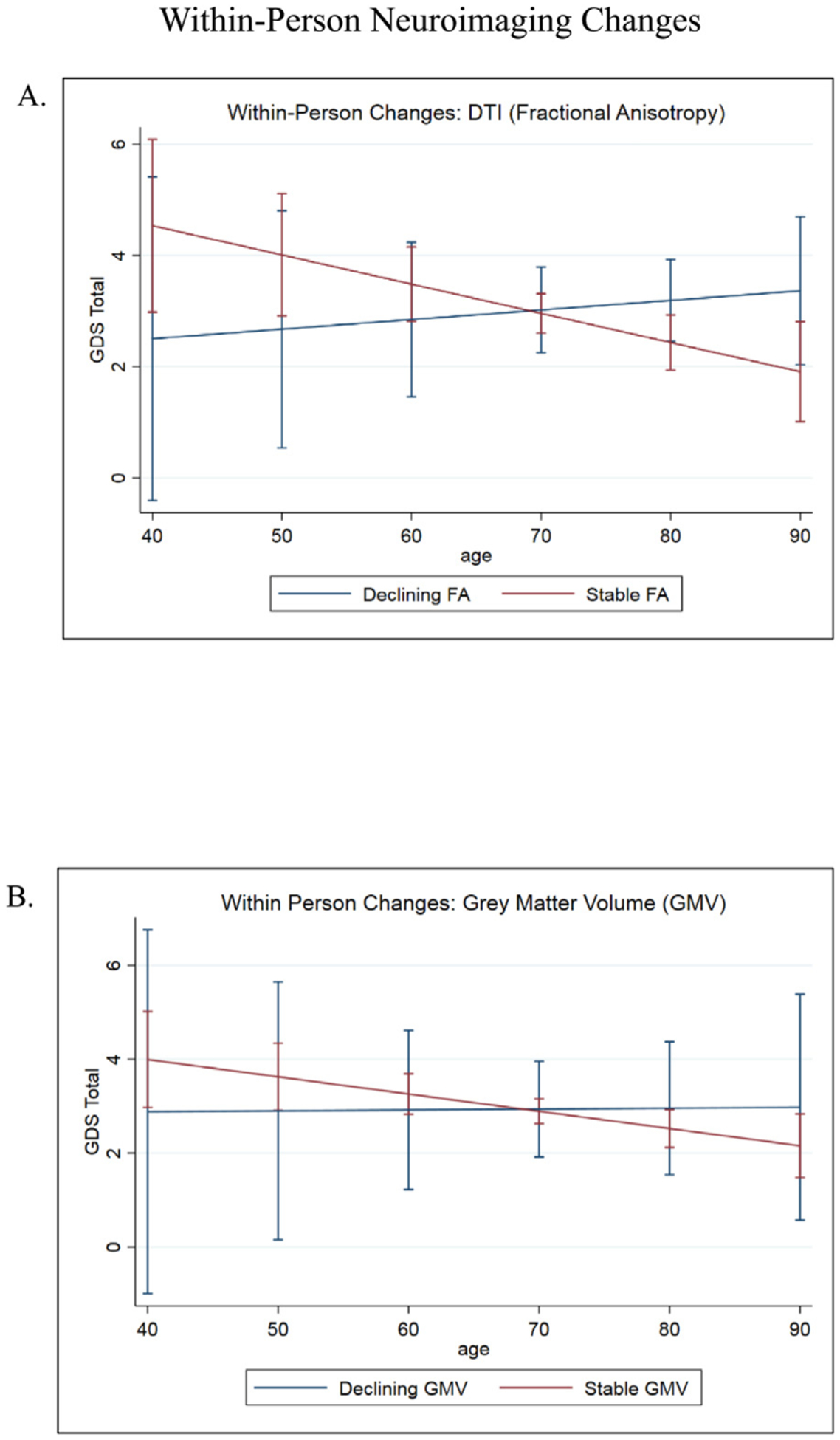
FIGURE 2.

[A] FA interacted with age to predict changes in GDS over time. In those with stable FA values, mood symptoms continued to improve over time. Mood symptoms worsened in those with declining FA values. Error bars indicate 95% CIs. [B] No significant associations between gray matter regions of interest, age, and mood. GM ROIs included medial temporal lobes, dorsolateral prefrontal cortex, amygdala, subcortical network, salience network, and executive network. Error bars indicate 95% CIs.
