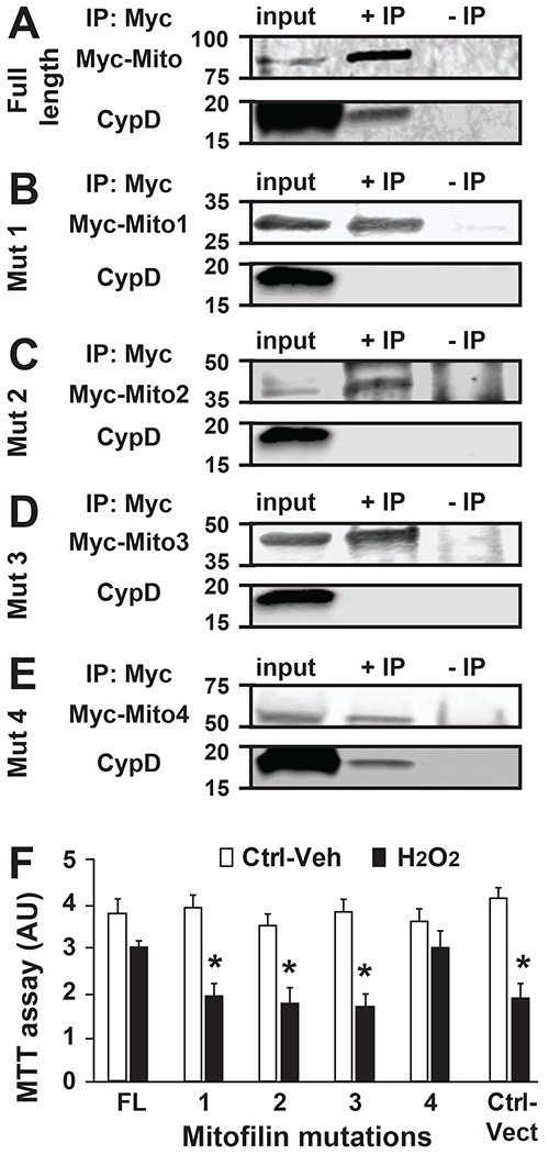
Figure 7. Mitofilin binds to CypD via c-terminal.

A-E. Immunoblots showing the C-terminal site in Mitofilin that bind to CypD. Mitofilin-mutated plasmids (1 to 5) were transfected in HEK293 cells and immunoprecipitation performed with Mitofilin-inserted-Myc in whole-cell lysate was followed by Western blot with anti-CypD antibody that allowed detection of CypD only when IP were performed in cells transfected with full-length-Mitofilin-Myc and Mitofilin-mutation4-Myc plasmid (A and E). This result indicates that the Mitofilin site that binds to CypD is located on the C-terminal that faces mitochondrial matrix IP: immunoprecipitation; IB: Western blot (immunoblot). F. Graph showing the level of cell death measure by MTT assay in HEK293 cells transfected with Mitofilin mutants as well as the full-length and treated with vehicle and the cytotoxic agent H2O2. Note that after treatment with H2O2, the cell death was reduced when the Mitofilin-CypD link was not disrupted (FL and 4) compared to plasmids (1, 2, and 3) and control vector. This observation suggests that the Mitofilin-CypD interaction in the IMM have a protective role against cell death. Values are expressed as mean±SEM; *P<0.05 full length (FL)-transfected plasmid (control) (n= 3/group triplicated in each). IP: immunoprecipitation; IB: Western blot (immunoblot).
