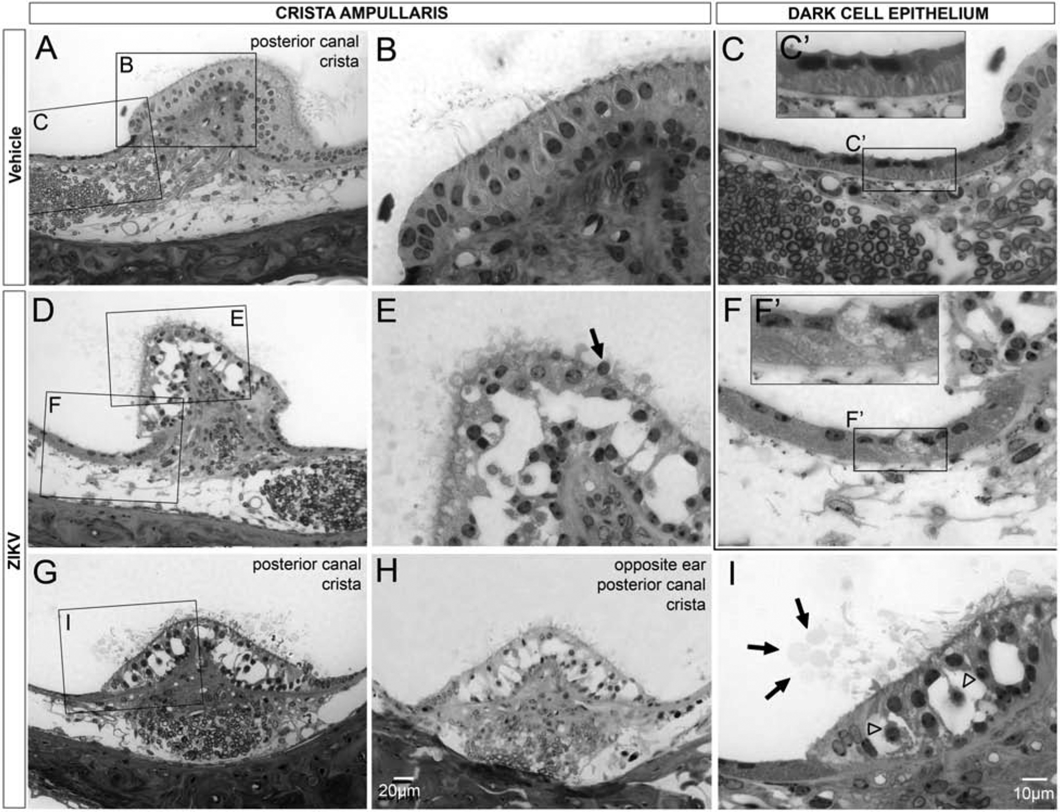
Figure 11.

Postnatal ZIKV infection of Ifnar1−/− mice causes damage to the epithelium of the crista ampullaris. The epithelium of the posterior canal crista shows normal integrity, with neighboring cells closely apposed, in vehicle treated mice (A, B). Vacuolation of the posterior canal crista epithelium is seen after ZKV infection (D, G, H; higher magnification E, I). Vacuolization the posterior canal crista occurs bilaterally (G, H). Cell shrinkage (open arrowheads, I), nuclear extrusion (arrow, E) and presence of vesicles above the apical surface of the crista epithelium (arrows, I) were all observed in the region of the crista ampullaris in ZIKV infected mice. The dark cell associated epithelium reveals normal integrity with striations beneath the apically located nuclei in vehicle-treated mice (C, C’). In ZIKV infected mice, the epithelium associated with the dark cells contain numerous vesicles that obliterate the normal striations and there is also loss of integrity of the apical surface (F, F’). Insets show higher magnification views of the dark cell epithelium (C’, F’). Scale bar for A, D, G, in H. Scale bar for B, C, E, F, in I.
